
|
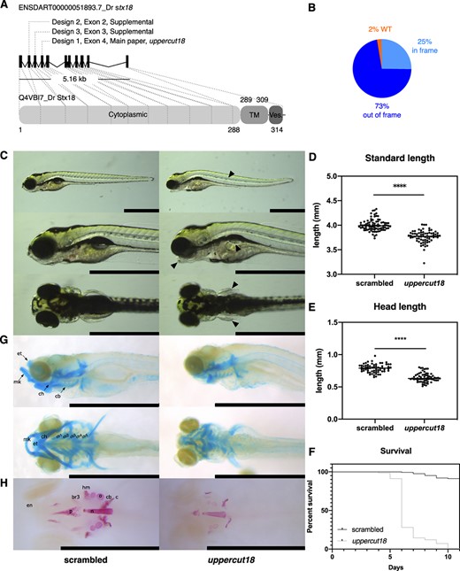
Fig. 2 Generation and characterization of mosaic stx18 zebrafish. (A) Zebrafish stx18 comprises 11 exons, is 5.16 kb long, and maps to chromosome 4. The stx18 protein (Q4VBI7) has similar domains as its human counterpart. The location of the CRISPR/Cas9 target sites are indicated and are located in exons 2 (design 2–validation purposes), 3 (design 3–validation purposes), and 4 (design 1–main article). (B) The majority of generated indels (98% of alleles) in the mosaic uppercut18 zebrafish model (“uppercut18 crispants”) result in a premature termination codon (PTC, 73% out‐of‐frame). (C) Uppercut18 zebrafish have a curved notochord (arrowhead), display cranial distortions (with severely deformed lower jaw, arrowhead), and have malformed pectoral fins (arrowheads) and an underdeveloped swim bladder that is not inflated (highlighted with a dotted white line and arrowhead). (D) Standard length of scrambled (n = 96) and uppercut18 (n = 86) zebrafish. The mean of data points is indicated, and an unpaired t test was used to determine significance. Each data point represents the measurement from 1 zebrafish, with highlighted median value (line). Two‐tailed unpaired t test; ****p < 0.0001. (E) Head length of scrambled (n = 64) and uppercut18 (n = 64) zebrafish. The mean of data points is indicated, and an unpaired t test was used to determine significance. Each data point represents the measurement from 1 zebrafish, with highlighted median value (line). Two‐tailed unpaired t test; ****p < 0.0001. (F) Survival curve of scrambled (n = 125) and uppercut18 (n = 127) zebrafish. All uppercut18 zebrafish died by 11 dpf. (G) Lateral (top) and ventral (bottom) views of Alcian blue–stained uppercut18 zebrafish reveal malformed Meckel's cartilage (mk), a smaller ethmoid plate (em), malformed ceratohyals (ch) and ceratobranchial pairs 1 to 5 (cb), and malformed pectoral fins (arrows). (H) Ventral view of Alizarin red–stained cranial skeleton reveals a generalized delay in ossification of the intramembranous and endochondral skeleton (p = parasphenoid; n = notochord; c = cleithrum; cb = ceratobranchial 5; o = opercle; hm = hyomandibular; br3 = branchiostegal ray 3) with absence of the entopterygoid (en). Images in C, G, and H are oriented to the left; scale bar = 1 mm. For C, G, and H, 20 5‐dpf scrambled and crispant zebrafish were analyzed and representative samples are shown.