
Image
|
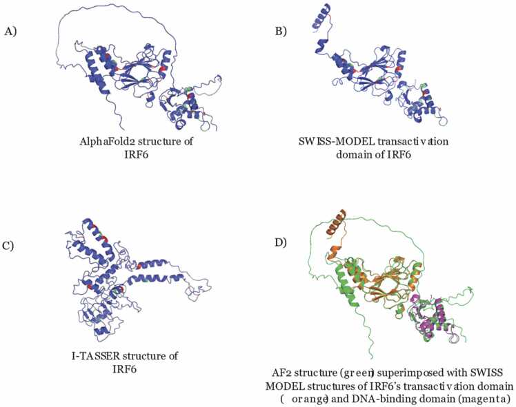
Figure Caption
Fig. 5
Predicted Protein Structures from different programs These images show (A) the AlphaFold2 structure prediction of IRF6 (residues 1–467), (B) the SWISS-MODEL structure prediction of the transactivation domain of IRF6 (residues 214–445), and (C) the I-TASSER structure prediction of IRF6 (residues 1–467) of highest confidence. Green residues were benign and red residues were pathogenic according to functional analysis. (D) The two SWISS-MODEL structures of the IRF6 transactivation and DNA-binding domains (residues 9–122) were superimposed onto the AlphaFold2 predicted structure.
Acknowledgments
This image is the copyrighted work of the attributed author or publisher, and
ZFIN has permission only to display this image to its users.
Additional permissions should be obtained from the applicable author or publisher of the image.
Full text @ Comput Struct Biotechnol J