
|
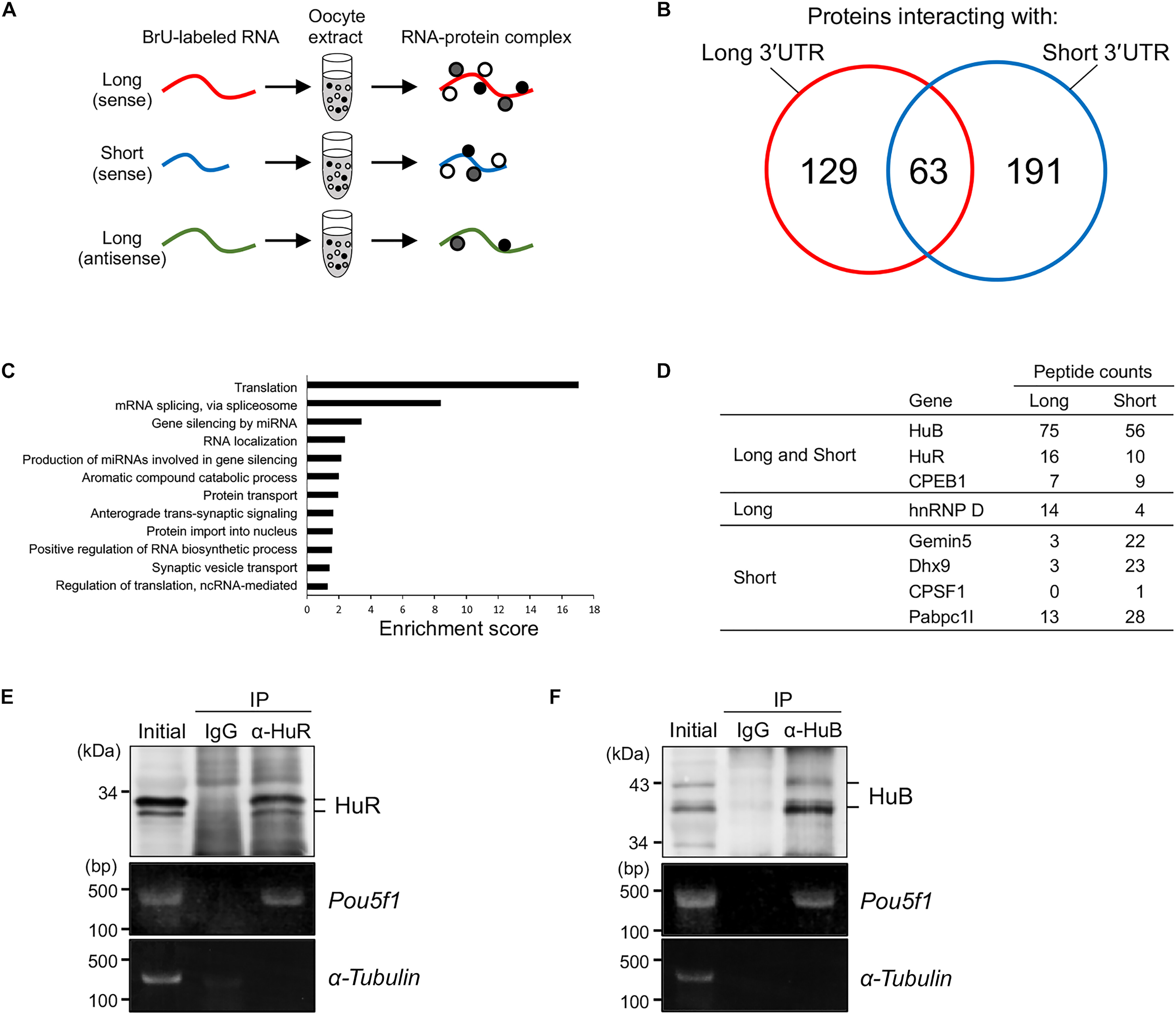
Fig. 6 Isolation of proteins binding to the long and/or short pou5f3 3′UTRs. (A) A schematic diagram of the isolation of proteins specifically binding to in vitro–synthesized pou5f3 long or short sense RNAs or long antisense RNA. (B) Venn diagram depicts the number of proteins isolated as proteins interacting with long (red) and short (blue) 3′UTR sequences of pou5f3 and the number of proteins binding to both 3′UTRs. (C) GO analysis of genes enriched in proteins isolated as proteins interacting with pou5f3 3′UTR sequences. (D) Representations of proteins isolated in the RNA pull-down assay. (E and F) IP/RT-PCR analysis of HuR and HuB with Pou5f1 mRNA. Top: Immunoblotting of mouse ovary extracts before IP (initial) and IP with control immunoglobulin G (IgG) or anti-HuR (α-HuR) (E) and anti-HuB (α-HuB) (F) antibodies. Bottom: Semiquantitative RT-PCR amplification of Pou5f1 and α-tubulin transcripts. Similar results were obtained from two independent experiments. BrU, bromouridine; ncRNA, noncoding RNA.