
|
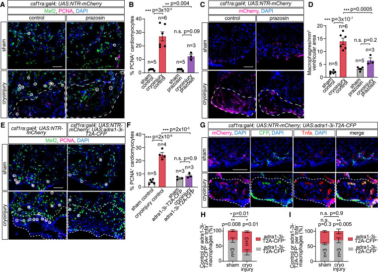
Fig. 3 Adra1 signaling regulates macrophage phenotypes and cardiomyocyte proliferation during inflammatory/scar resolution in injured hearts (A and B) Adult csf1ra:Gal4; UAS:NTR-mCherry hearts following sham operation or cryoinjury with control solution or prazosin treatment for 7 days. Immunofluorescent staining of Mef2 and PCNA labels cardiomyocytes and proliferating cells, respectively, in the heart cryosections (A). Proliferating cardiomyocytes (Mef2+/PCNA+) in the proximity of injury border zones, indicated by white circles (A) and quantified as percentages of cardiomyocytes within the observed area (B). (C and D) Macrophages, labeled by mCherry immunostaining, in cryosections of hearts of adult csf1ra:Gal4; UAS:NTR-mCherry fish subjected to sham operation or cryoinjury, treated with control solution or prazosin for 7 days (C), and quantified as cell numbers per 1 mm2 ventricular area (D). (E and F) Proliferating cardiomyocytes in adult csf1ra:Gal4; UAS:NTR-mCherry (control) and csf1ra:Gal4; UAS:NTR-mCherry; UAS:adra1-3i-T2A-CFP (adra1-3i-T2A-CFP) animals labeled by immunofluorescent staining for Mef2 and PCNA, indicated by white circles (E) and quantified as percentages of PCNA+ cardiomyocytes within the observed area (F). (G) Sham and cryoinjured csf1ra:Gal4; UAS:NTR-mCherry; UAS:adra1-3i-T2A-CFP heart sections, immunostained for mCherry (all macrophages), CFP (adra1-3i-T2A-CFP+ macrophages), and Tnfa at 7 dpi. (H and I) Segmented bar graphs showing distribution of adra1-3i-T2A-CFP– (control) and adra1-3i-T2A-CFP+ macrophages within the pro-inflammatory (tnfa+) (H) and non-inflammatory (tnfa−) (I) pools present in sham-operated and cryoinjured hearts at 7 dpi. DAPI staining labeled all nuclei. Lesioned areas are outlined by white dashed lines. All scale bars: 50 μm. Data are presented as mean ± SEM, with data points of individual animals. n denotes number of animals measured in each group. ∗ p < 0.05, ∗∗ p < 0.01, ∗∗∗ p < 0.001; n.s. not significant, two-tailed t test.
Reprinted from Developmental Cell, 58(22), Apaydin, O., Altaikyzy, A., Filosa, A., Sawamiphak, S., Alpha-1 adrenergic signaling drives cardiac regeneration via extracellular matrix remodeling transcriptional program in zebrafish macrophages, 2460-2476.e7, Copyright (2023) with permission from Elsevier. Full text @ Dev. Cell