IMAGE

Fig. 5

ID
ZDB-IMAGE-240212-33
Source
Figures for Kim et al., 2024
Image
Figure Caption

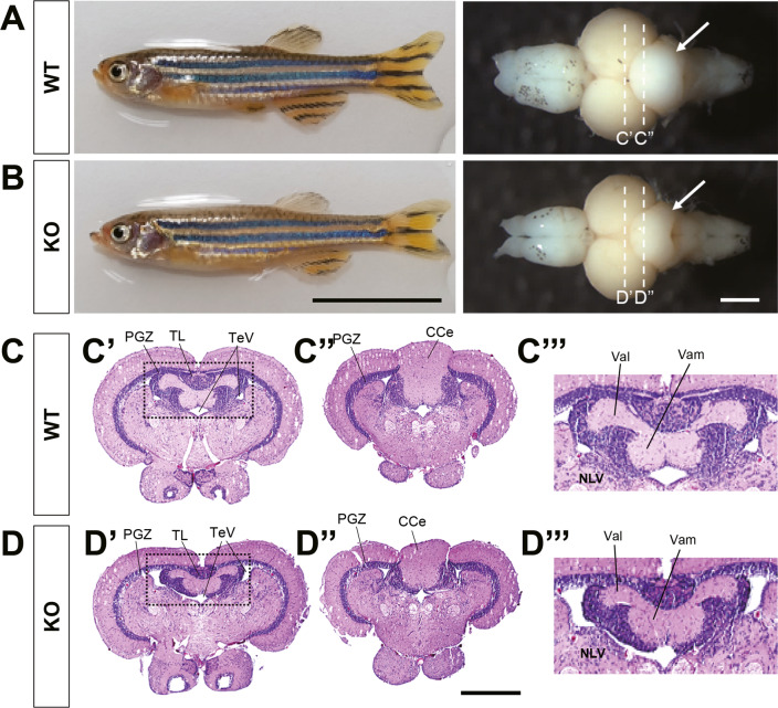
Fig. 5 Anatomical and histological analysis of adult brains.

A, B Representative images of znf536 KO adult zebrafish at 11 mpf and their isolated brains. Arrows indicate the reduced size of the cerebellum in KO zebrafish. n = 7 for WT and n = 6 for KO. Scale bar: 10 mm (left panels) and 500 µm (right panels). C, D Brain section images at positions indicated by dashed lines in A, B. Serial sections were carefully examined to compare relative planes. C”’, D”’ Magnification of marked areas in C’, D’ (dashed box). ImageJ analysis C’, D’ showed that the relative size of valvular cerebelli of znf536 KO zebrafish (average 7.8% of the whole brain, n = 4) was reduced, compared to that of WT zebrafish (average 9.6%, n = 3). CCe corpus cerebelli, NLV nucleus lateralis valvulae, PGZ periventricular gray zone of optic tectum, TeV tectal ventricle, TL torus longitudinalis, Val lateral division of valvular cerebelli, Vam medial division of valvular cerebelli. Scale bar: 500 µm.

Acknowledgments
This image is the copyrighted work of the attributed author or publisher, and ZFIN has permission only to display this image to its users. Additional permissions should be obtained from the applicable author or publisher of the image. Full text @ Transl Psychiatry