
Fig. 6
- ID
- ZDB-IMAGE-240206-31
- Publication
- Birdal et al., 2023 - Expression of taste sentinels, T1R, T2R, and PLCß2, on the passageway for olfactory signals in zebrafish
- All Figures
- Figures for Birdal et al., 2023
|
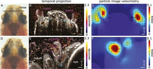
Fig. 6 Directional fluid flow in the nostrils of adult zebrafish. (A, D): Dorsal view of an adult zebrafish head with the nose highlighted. The black bars indicate the location of the OCT recordings. Note that (A) represents a coronal and (D) a sagittal view of the zebrafish nose. (B, E): Projection of a 100 frames sequence (equivalent to 2.3 s) of OCT images of a deeply anesthetized adult zebrafish. Time 0 is indicated in dark magenta and time 2.3 s is indicated in yellow as shown in the false color scale. The anatomy of the nose, including the nostril and olfactory epithelium (OE), is indicated with dashed lines. Note the rapid movement of particles in the area surrounding the nostrils. Since the animal is deeply anesthetized, there is a reduced respiratory rate and little movement of particles around the mouth. (C, F): Particle image velocimetry analysis reveals the directionality and velocity of the fluid flow around the head of a deeply anesthetized zebrafish. Note that the highest velocity occurs at the inlet of the nostrils. Scale bar: 50 µm.