
|
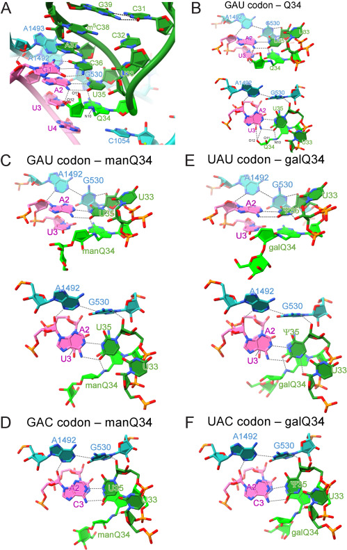
Fig. S7 Cryo-EM analyses of codon-anticodon interactions of human tRNAs at the A-site of E. coli 70S ribosome complexes, related to Figure 6 (A) Close-up view of the codon-anticodon interaction at the A-site of the ribosome with human tRNAAsp bearing Q34 (green) and GAU codon (magenta) (PDB: 7Y7D ). 16S rRNA is colored cyan. Hydrogen bonds are shown as dotted lines. (B, C, and E) Cryo-EM structure of the codon-anticodon interaction at the A-site between human tRNAAsp bearing Q34 (green) and the GAU codon (PDB: 7Y7D ) (B), human tRNAAsp bearing manQ34 (green) and the GAU codon (PDB: 7Y7E) (C), and human tRNATyr bearing galQ34 (green) and the UAU codon (PDB: 7Y7G ) (E), viewed from the major groove side (upper) and from the upper side (lower). Hydrogen bonds are shown as dotted lines. (D and F) Cryo-EM structure of the codon-anticodon interaction at the A-site between human tRNAAsp bearing manQ34 (green) and the GAC codon (PDB: 7Y7F ) (D), and human tRNATyr bearing galQ34 (green) and the UAC codon (PDB: 7Y7H) (F), viewed from the upper side. Hydrogen bonds are shown as dotted lines.
Reprinted from Cell, 186(25), Zhao, X., Ma, D., Ishiguro, K., Saito, H., Akichika, S., Matsuzawa, I., Mito, M., Irie, T., Ishibashi, K., Wakabayashi, K., Sakaguchi, Y., Yokoyama, T., Mishima, Y., Shirouzu, M., Iwasaki, S., Suzuki, T., Suzuki, T., Glycosylated queuosines in tRNAs optimize translational rate and post-embryonic growth, 5517-5535.e24, Copyright (2023) with permission from Elsevier. Full text @ Cell