
|
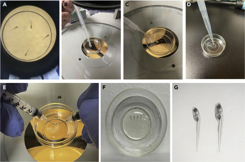
Fig. 3 Embed larvae in LMA for imaging (A) Transfer the larvae into a 35 mm Glass Bottom Dish. (B) After anesthesia, remove most of the medium with a transfer pipette. (C) Remove the residual medium using a transfer pipette with a 10 μL pipette tip attached. (D) Add 750 μL of the pre-heated 1.2% LMA at the center of glass bottom using a P1000 pipette. (E) Position the larvae on their right side using two insulin syringe needles. (F) Add 750 μL 0.3x Danieau’s solution (containing Tricaine) at the periphery of the dish after the LMA solidifies to submerge the gel. (G) Check the larvae under a stereo microscope.