IMAGE

Figure 5

ID
ZDB-IMAGE-240129-258
Source
Figures for Ng et al., 2024
Image
Figure Caption

Figure 5

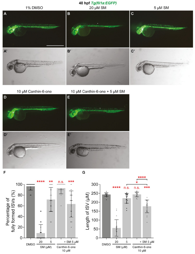
Canthin-6-one synergises with VEGFR inhibitor sunitinib malate. (AE’) Lateral fluorescent (AE) and trans-light images (AE’) of 48 hpf Tg(fli1a:EGFP) embryos treated with either 1% DMSO (A,A’), 20 µM sunitinib malate (SM, B,B’), 5 µM SM (C,C’), 10 µM canthin-6-one (D,D’), or co-treatment of 5 µM SM and 10 µM canthin-6-one (E,E’). White asterisks indicate the absence of intersegmental vessels (ISVs). (F,G) Quantification of the percentage of fully formed ISVs (F) or length of ISVs (G) in 48 hpf Tg(fli1a:EGFP) embryos treated with either 1% DMSO (n = 23), 20 µM SM (n = 22), 5 µM SM (n = 23), 10 µM canthin-6-one (n = 17), or co-treatment of 5 µM SM and 10 µM canthin-6-one (n = 21). Statistical test: Kruskal-Wallis test was conducted for graphs (F,G). p ≤ 0.0001 (****), p ≤ 0.001 (***), p ≤ 0.01 (**), p ≤ 0.05 (*), p > 0.05 (not significant, n.s.). Scale bar: 1 mm (A).

Acknowledgments
This image is the copyrighted work of the attributed author or publisher, and ZFIN has permission only to display this image to its users. Additional permissions should be obtained from the applicable author or publisher of the image. Full text @ Pharmaceuticals (Basel)