IMAGE

Fig. 8.

ID
ZDB-IMAGE-240129-252
Source
Figures for Olson et al., 2024
Image
Figure Caption

Fig. 8.

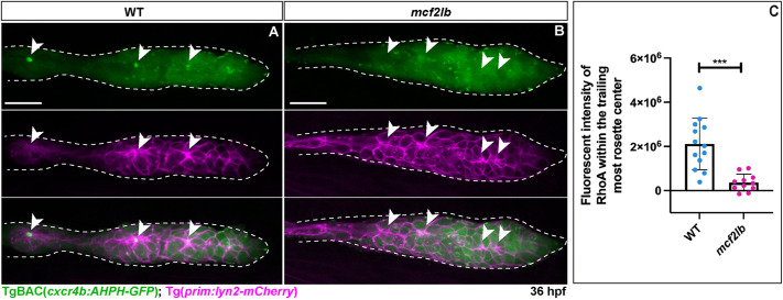
RhoA signal is lost at rosette centers in mcf2lb mutants. (A,B) Fluorescent signal from the RhoA sensor line TgBAC(cxcr4b:AHPH-GFP) at 36 hpf in WT (A) and the mcf2lb mutant (B). Tg(prim:lyn2-mCherry) marks cell membranes in the migrating pLLP. Dashed lines mark the pLLP. Arrowheads indicate rosette centers and points. Note the absence of RhoA signal at the rosette centers and points in the mcf2lb mutant pLLP. (C) Quantification of RhoA signal at the center of the trailing-most rosette (point) within the migrating pLLP in WT (n=13) and mcf2lb mutants (n=10). ***P<0.001 (unpaired two-tailed t-test). Error bars are s.d. Scale bars: 20 μm.

Acknowledgments
This image is the copyrighted work of the attributed author or publisher, and ZFIN has permission only to display this image to its users. Additional permissions should be obtained from the applicable author or publisher of the image. Full text @ Development