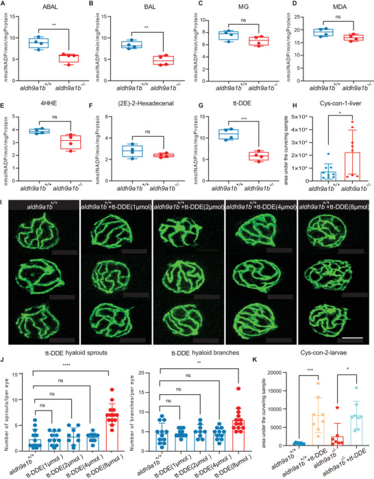
IMAGE

Figure 3

ID
ZDB-IMAGE-240129-205
Source
Figures for Qian et al., 2023
Image
Figure Caption

Figure 3

The link between tt‐DDE, Aldh9a1b, and angiogenesis (A‐G) Aldh enzyme activity was significantly decreased using substrate ABAL (A), BAL (B), and tt‐DDE (G), but unaltered with substrate MG (C), MDA (D), 4HHE (E) and 2(E)−2‐hexadecenal (F) in aldh9a1b−/‐ larvae at 5dpf. Each data point represented 120 larvae per clutch, n = 4. H) Cys‐con‐1 was significantly changed in aldh9a1b−/− livers. n = 9/8. I) Representative confocal images of hyaloid vasculature showed vascular alterations in aldh9a1b+/+ larvae treated with 0–8 µmol tt‐DDE at 5dpf. White scale bar = 50 µm. J) Quantification of increased hyaloid branch points and sprouts formation in 8 µmol tt‐DDE treated aldh9a1b+/+ zebrafish larvae, while no significant change in 1, 2, and 4 µmol tt‐DDE treatments were observed. One data point means one hyaloid per larva. K) Cys‐con2 was significantly increased in tt‐DDE‐treated larvae. n = 8/7. The bars indicate mean ± SD values. Statistical analysis was performed by Student's t‐test and one‐way ANOVA. ns = not significant, *p < 0.05, **p < 0.01, ***p < 0.001, ****p < 0.0001.

Acknowledgments
This image is the copyrighted work of the attributed author or publisher, and ZFIN has permission only to display this image to its users. Additional permissions should be obtained from the applicable author or publisher of the image. Full text @ Adv Sci (Weinh)