Figure Caption
Fig 2
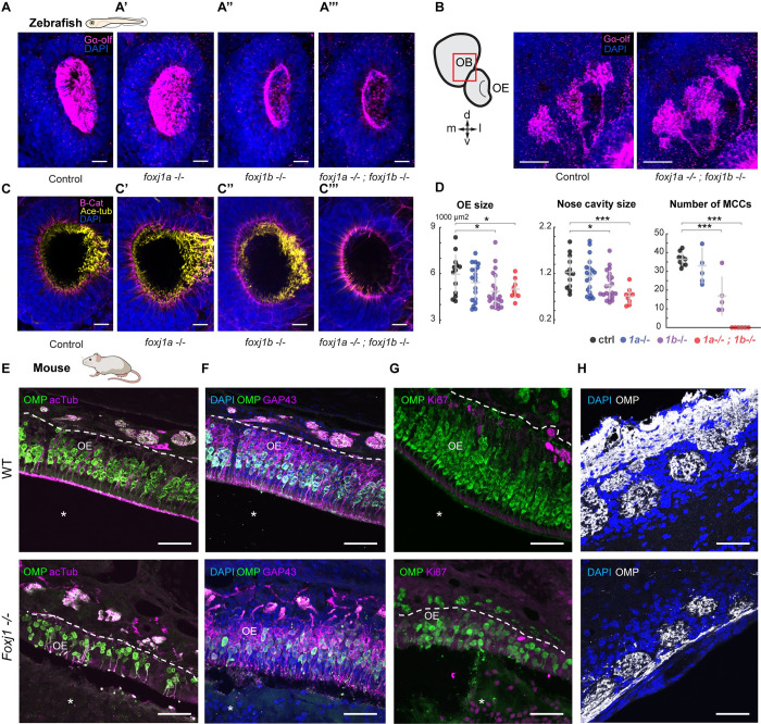
Foxj1 regulates olfactory cilia biogenesis in zebrafish and OE establishment in mice.
(A) Immunostaining with anti-Gαolf antibody marking olfactory cilia (magenta) in foxj1a,b mutant zebrafish larvae at 4 dpf. Nuclei marked with DAPI (blue). foxj1a mutants showed no observable effect on the formation of olfactory cilia (n = 3). foxj1b mutants showed reduced olfactory cilia (n = 3). foxj1a/b double mutants showed severe loss of olfactory cilia (n = 3). (B, B’) Innervation pattern of ciliated OSNs labeled with Gαolf (magenta) in control (n = 3) and foxj1a/b double mutant (n = 5) showed no significant effect in the formation of olfactory glomeruli at 4 dpf. Nuclei marked with DAPI (blue). (C) foxj1a/b double mutant showing severe reduction of motile cilia number in comparison to the foxj1a mutants, foxj1b mutants and control, as shown by immunostaining with anti-acetylated-tubulin for marking cilia (yellow) and beta-catenin for marking cell borders (magenta) at 4 dpf. (nControl = 13, nfoxj1a−/− = 20, nfoxj1b−/− = 22, nfoxj1a−/−; foxj1b−/− = 9). Nuclei are marked with DAPI (blue). (D) Significant decrease in the size of nose and nasal cavity, and number of MCCs in foxj1b and foxj1a/b double mutant embryos. (For nose size, nControl = 13, nfoxj1a−/− = 20, nfoxj1b−/− = 22, nfoxj1a−/−; foxj1b−/− = 9. For number of MCCs, nControl = 9, nfoxj1a−/− = 5, nfoxj1b−/− = 5, nfoxj1a−/−; foxj1b−/− = 6). (E) The OE of P21 mouse was stained for OMP (green), a mature OSN marker. Apical layer, composed of mucus and cilia, is strongly labeled for acetylated α-tubulin (magenta) in control (top, arrow). Mature OSNs (OMP, green) at the same animal age of 21 day were fewer in number and disorganized within the OE of the Foxj1−/− mouse (bottom), (10,630 ± 923 cells per mm2, n = 8, WT; 3,106 ± 714, n = 7, KO, 3 mice, p = 0.0006). Compared to the WT, thickness of the OE was significantly reduced in the Foxj1−/− mutant (79.68 ± 4.31 μm, n = 34, 3 mice, WT; 45.48 ± 2.15 μm, n = 30, 3 mice, p < 0.0001, KO). (F) Immature OSNs (GAP43, magenta) were located below the layer of mature OSNs (OMP, green) as shown in a control mouse (top). Immature OSNs (GAP43, magenta) lost their orientation relative to mature OSNs and were fewer in numbers within the OE of the Foxj1−/− mutant (bottom) as compared to control (6,339 ± 1,015 cells per mm2, n = 8, WT; 2,008 ± 481, n = 7, KO, 3 mice, p = 0.0012). The nasal cavity (E-G, asterisks) was unobstructed in the WT, whereas in the Foxj1−/− animals, it was completely filled with DAPI and Ki67-positive cells. (G) Proliferating cells expressing Ki67 (magenta) were mostly comprised of basal progenitor cells lining lamina propria. Fewer proliferating cells (Ki67, magenta) were present in the OE of the Foxj1−/− mouse (bottom) as compared to the control (top) (2,362 ± 321 cells per mm2, n = 9, WT; 502 ± 72, n = 9, KO, 3 mice, p < 0.0001). (H) Images showing the OB having oval-shaped glomeruli (top) filled with the axonal projections of OSNs (OMP, white) in control (top). Reduced intensity of OMP immunostaining in the Foxj1−/− mouse (bottom) as compared to the control (top) (352.5 ± 26.7 a.u., n = 21, WT; 266.6 ± 17.5 a.u., n = 44, KO; 3 mice, p = 0.0133). In the Foxj1−/− mouse, glomeruli overall were less developed and had smaller perimeter than in the WT (2,558 ± 368 μm, n = 21, 2 mice, WT; 1,331 ± 306 μm, n = 44, 2 mice, KO; 3 mice, p < 0.0001). Scale bars: 10 μm (A-D), 50 μm (E-H). Raw data files are available in Mendeley Data (https://data.mendeley.com/datasets/2pn963jn6y). dpf, days post fertilization; KO, knockout; MCC, motile multiciliated cell; OB, olfactory bulb; OE, olfactory epithelium; OMP, olfactory marker protein; OSN, olfactory sensory neuron; WT, wild type.
Acknowledgments
This image is the copyrighted work of the attributed author or publisher, and
ZFIN has permission only to display this image to its users.
Additional permissions should be obtained from the applicable author or publisher of the image.
Full text @ PLoS Biol.