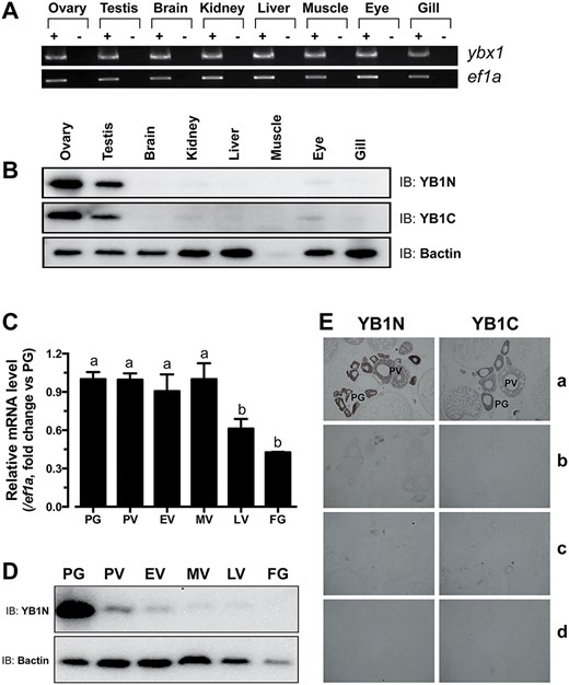
Fig. 3 Spatiotemporal expression of Ybx1 mRNA and protein in different tissues and ovarian follicles. (A) Semi-quantitative RT-PCR analysis of ybx1 and ef1a expression in different tissues. +, RT with M-MLV; −, RT without M-MLV. (B) Western blot detection of Ybx1 in different tissues. YB1N, anti-N-terminal antibody; YB1C, anti-C-terminal antibody; Bactin, β-actin. (C) Real-time PCR analysis of ybx1 expression in the follicles of different stages. Housekeeping gene, ef1a, was used as control. Different letters indicate statistical significance (n = 3) (D) Western blot detection of Ybx1 protein in different stages of follicles. (E) Immunohistochemical detection of Ybx1 in follicles. a, with antibodies YB1N or YB1C; b, antibodies neutralized with the antigenic peptides; c, pre-immune sera; d, no antibodies.
Image
Figure Caption
Acknowledgments
This image is the copyrighted work of the attributed author or publisher, and
ZFIN has permission only to display this image to its users.
Additional permissions should be obtained from the applicable author or publisher of the image.
Full text @ Biol. Reprod.

