
Image
|
Figure Caption
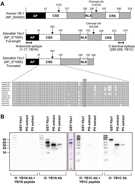
Fig. 2 Structure of zebrafish Ybx1 and preparation of anti-Ybx1 antibodies. (A) Domain structure of human YB-1 and zebrafish Ybx1 proteins. AP, AP-rich N-terminal; CSD, cold shock domain; NLS, nuclear localization signal; CRS, cytosol localization signal; YB1N, anti-N-terminal antibody; YB1C, anti-C-terminal antibody. (B) Western blot testing for antibody specificity. YB1N could detect a major band at 48 kDa and a minor band at 36 kDa (asterisk) in PG follicle extract, whereas the YB1C could detect the major band only.
Acknowledgments
This image is the copyrighted work of the attributed author or publisher, and
ZFIN has permission only to display this image to its users.
Additional permissions should be obtained from the applicable author or publisher of the image.
Full text @ Biol. Reprod.