
Fig. 3
- ID
- ZDB-IMAGE-240124-32
- Publication
- Rodríguez-Ruiz et al., 2023 - ZAKα/P38 kinase signaling pathway regulates hematopoiesis by activating the NLRP1 inflammasome
- All Figures
- Figures for Rodríguez-Ruiz et al., 2023
|
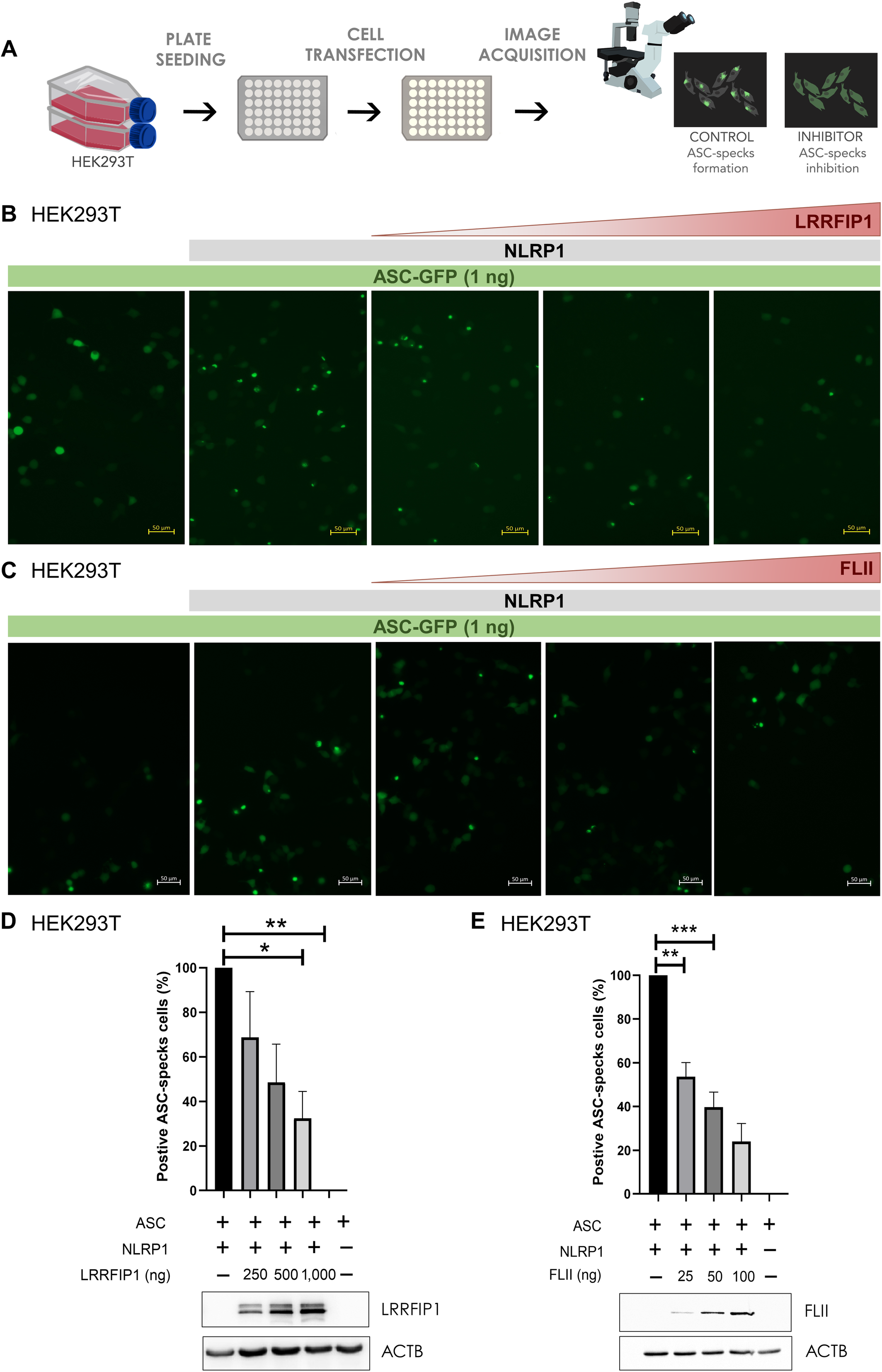
Fig. 3 LRRFIP1 and FLII negatively regulate human NLRP1 inflammasome activation A HEK293T cells were transfected with 300 ng NLRP1‐FLAG, 1 ng ASC‐GFP and the indicated concentrations of FLAG‐LRRFIP1 and FLAG‐FLII plasmids, and the formation of ASC specks was analyzed by fluorescence microscopy 24 h post‐transfection. B, C Representative images of ASC specks. D, E Number of positive ASC specks in cells co‐transfected with LRRFIP1 (D) and FLII (E). Expression of LRRFIP and FLII was confirmed by Western blot using anti‐FLAG and anti‐ACTB antibodies. Data are shown as the means ± SEM (n = 95–156 cells). P values were calculated using one‐way ANOVA and Tukey's multiple range test. *P < 0.05; **P < 0.01; ***P < 0.001.