
Fig. 6
- ID
- ZDB-IMAGE-240112-40
- Publication
- Mahony et al., 2023 - Lineage skewing and genome instability underlie marrow failure in a zebrafish model of GATA2 deficiency
- All Figures
- Figures for Mahony et al., 2023
|
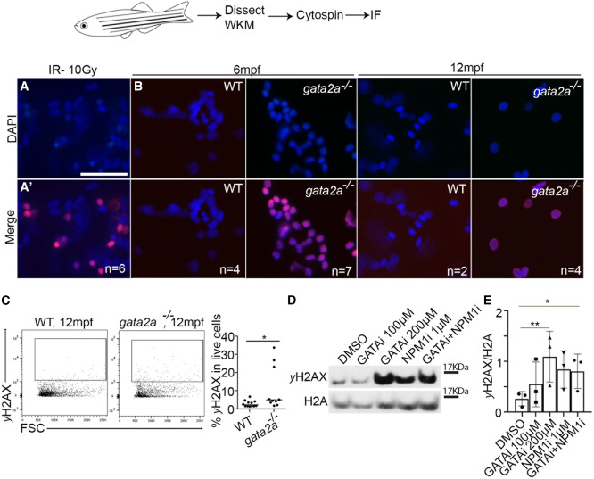
Fig. 6 Loss of gata2a leads to increased DNA damage and mutation burden in WKM of gata2a−/− MUTs (A and A′) Control irradiation performed at 10 Gy on WKM cells and immunofluorescence against DAPI and γH2AX. Scale, 25 μm. (B) WKM from 6 and 12 mpf WT or gata2a−/− adults and immunofluorescence against DAPI and γH2AX. Imaging is at 40× magnification. (C) Flow cytometry analysis of γH2AX expression in 12 mpf WKM. p = 0.0296, WT: n = 12 (biological replicates), gata2a−/−: n = 10 (biological replicates). (D) Western blot of γH2AX and H2A (loading control) in HPC7 cells after 1 μM NSC348884 (NPM1 inhibitor43) or 100/200 μM pyrrothiogatain (GATA inhibitor49) treatment for 24 h. (E) Quantification of western blot data, n = 3/condition (biological triplicates). ∗p < 0.05, determined by using an unpaired, two-tailed t test in (C). ∗∗p < 0.01 and ∗p < 0.05, determined by Friedman test in (E). See also Figure S6.