
|
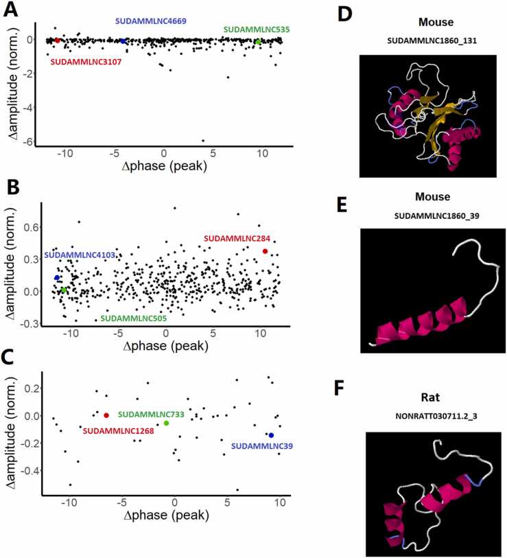
Fig. 8
Visualization of changes of the phases and amplitudes of loss-of-rhythmicity, gain-of-rhythmicity, and rhythmicity-maintaining lncRNAs between control and desynchronized conditions, and 3-dimensional structures of the conserved lncRNA-encoded peptides. (A-C) Changes in the phases and amplitudes of loss-of-rhythmicity l mouse ncRNAs (A) and gain-of-rhythmicity mouse lncRNAs in the desynchronized condition (B), and rhythmicity-maintaining mouse lncRNAs between control and desynchronized conditions (C). (D-F) 3D models of peptides encoded by two mouse testicular lncRNAs (SUDAMMLNC1860_131, and SUDAMMLNC1860_39) (D, E) conserved with one rat lncRNA (NONRATT030711.2_3) (F). The predicted 3D models of the conserved lncRNA-encoded peptides revealed the presence of α-helix (pink or purple motif structure), β-strand (yellow layered band), and random coils (white or blue thread) with the known domains from Protein Data Bank, such as D (1g3kA), E (2axtL), and F (1g5cA).