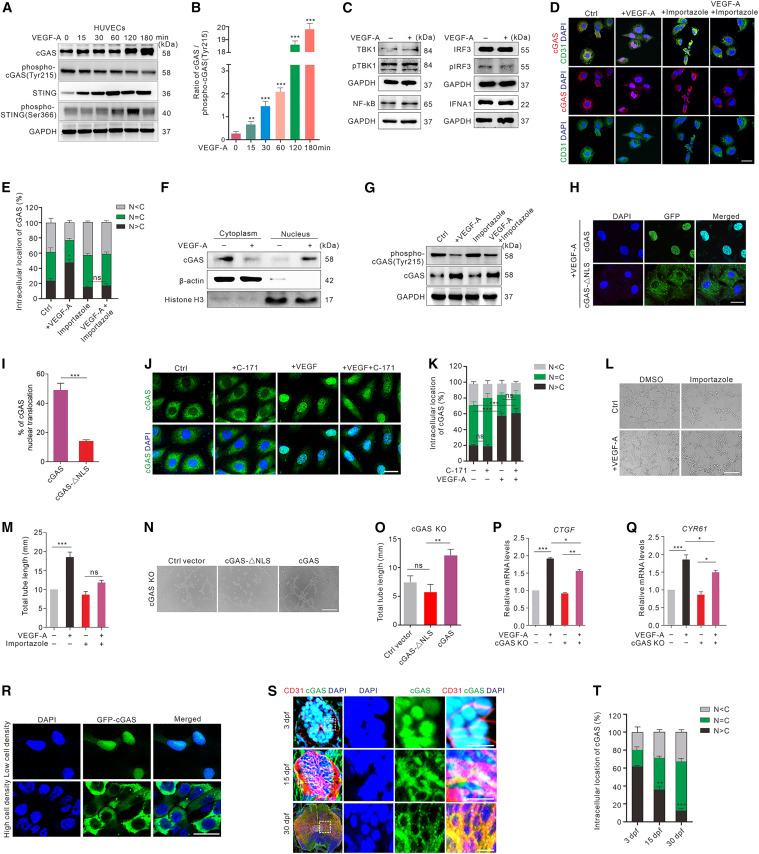
Fig. 3 cGAS nuclear translocation is involved in VEGF-A-mediated angiogenesis (A and B) Western blot (A) and quantification (B) of the dephosphorylation of cGAS and STING in HUVECs after VEGF-A stimulation at different time points. (C) The expression of IFNA1, NF-κB, IRF3, pIRF3, TBK1, and pTBK1 in HUVECs with or without VEGF-A stimulation (n = 3 replicates). (D and E) Immunofluorescence (D) and quantification (E) of cGAS (red) intracellular localization in HUVECs treated with or without VEGF-A and/or importazole (n = 12 random FOV/group). Scale bar, 20 μm. (F) The expression of cGAS in nuclear and cytoplasmic fractions in HUVECs with or without VEGF-A stimulation (n = 3 replicates). (G) cGAS dephosphorylation in HUVECs treated with or without VEGF-A and/or importazole. (H and I) Representative images (H) and quantification (I) of the distributions of GFP-labeled cGAS or cGAS with NLS-directed mutagenesis in GFP-FLAG-cGAS-ΔNLS or GFP-FLAG-cGAS vector-transfected cGAS KO HUVECs after VEGF-A stimulation (n = 12 random FOV/group). Scale bar, 20 μm. (J and K) Representative images (J) and quantification (K) of cGAS (green) intracellular localization in VEGF-A and/or C-171-treated HUVECs (n = 12 random FOV/group). Scale bar, 20 μm. (L and M) Tube formation (L) and quantification (M) in HUVECs with or without VEGF-A and/or importazole treatment (n = 3 replicates). Scale bar, 200 μm. (N and O) Tube formation (N) and quantification (O) in control vector-, GFP-FLAG-cGAS-ΔNLS-, or GFP-FLAG-cGAS-transfected cGAS KO HUVECs (n = 3 replicates). Scale bar, 200 μm. (P and Q) Relative mRNA levels of CTGF (P) and CYR61 (Q) in cGAS KO and normal HUVECs before and after 6-h VEGF-A treatment (n = 4 replicates). (R) Representative images of cGAS intracellular distributions in HUVECs stably expressing GFP-cGAS (green) with low- (40% confluence) or high-density (80% confluence). Scale bar, 20 μm. (S and T) Immunofluorescence (S) and quantification (T) of cGAS (green) intracellular localization at different developmental stages in SC of zebrafish (n = 5 replicates). Scale bars, 100 μm (lower magnification) and 20 μm (insets). Data are represented as means ± SEM. Student’s t test in (I), ANOVA in (B), (E), (K), (M), (O–Q), and (T). ∗p < 0.05; ∗∗p < 0.01; ∗∗∗p < 0.001; ns, no significant. C, cytosol; N, nucleus. See also Figures S2 and S3.
Image
Figure Caption
Acknowledgments
This image is the copyrighted work of the attributed author or publisher, and
ZFIN has permission only to display this image to its users.
Additional permissions should be obtained from the applicable author or publisher of the image.
Full text @ Cell Rep.

