
|
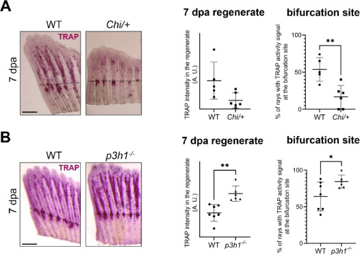
Fig. 9 Osteoclast activity in the regenerate and at the bifurcation site at 7 dpa in WT, Chi/+ and p3h1−/−. (A) TRAP staining of WT and Chi/+ caudal fin samples collected at 7 dpa (the amputation site is indicated by the black dotted line). While total TRAP activity in the regenerate was unchanged between WT and Chi/+, TRAP signal at the bifurcation site was significantly reduced in Chi/+ respect to WT (n ≥ 5 caudal fins for each genotype, as indicated by the dots). Scale bar: 500 µm. (B) TRAP staining of WT and p3h1−/− caudal fin samples collected at 7 dpa (the amputation site is indicated by the black dotted line). TRAP activity was significantly increased in p3h1−/− with respect to WT at this time point, with strong TRAP signal detected at the level of the bifurcations (n ≥ 6 caudal fins for each genotype, as indicated by the dots). Scale bar: 500 µm. * P < 0.05, ** P < 0.01.