IMAGE

Fig. 1.

ID
ZDB-IMAGE-231220-41
Source
Figures for Square et al., 2023
Image
Figure Caption

Fig. 1.

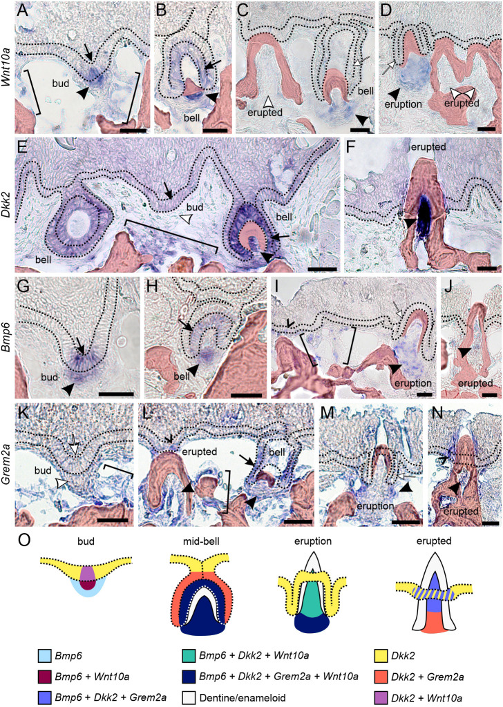
In situ hybridization reveals dynamic expression of Wnt10a, Dkk2, Bmp6 and Grem2a within and surrounding pharyngeal teeth in wild-type sticklebacks. (A-N) The basal-most layer of epithelium is flanked by black dotted lines in each image. Arrows mark dental epithelium and arrowheads mark dental mesenchyme, with black markers indicating detected expression and white markers indicating regions with no detected expression; brackets mark mesenchymal expression outside of tooth organs; carets mark the successional dental epithelium. Bony tissues are false-colored red. Tooth stages and genes are labeled in the figure. Each expression pattern presented was observed in at least n=3 individuals. (O) A diagram showing bud, mid-bell, eruption, and erupted teeth summarizing the expression domains observed. The dotted lines demarcate the basal-most layer of epithelium. The hashed domain in the erupted stage indicates that these genes are not always observed in the successional dental epithelium of an erupted tooth. Descriptions of the location of the successional dental epithelium can be found in Fig. 10 in this paper and figure 6 in Square et al. (2021). Scale bars: 20 μm.

Acknowledgments
This image is the copyrighted work of the attributed author or publisher, and ZFIN has permission only to display this image to its users. Additional permissions should be obtained from the applicable author or publisher of the image. Full text @ Development