
|
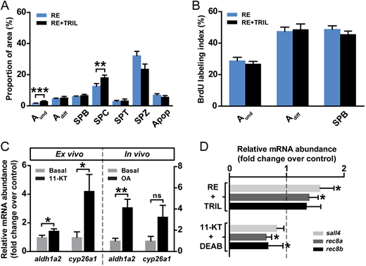
Fig. 6 The influence of steroids on the regulation of retinoid-modulated spermatogenesis. (A,B) Frequency of germ cell types and apoptotic cells (A) and BrdU labeling indices (B) in testicular explants cultured for 4 days in the presence of 10 µM RE or plus 25 µg/ml trilostane (TRIL). Data are mean±s.e.m. (n=6; **P<0.01, ***P<0.001). (C) Ex vivo and in vivo androgen effects on aldh1a2 and cyp26a1 expression. In the left panel, testis tissue was cultured for 4 days to study the effects of 11-KT (200 nM) on the mRNA abundance of RA-related enzymes compared with the control group. In the right panel, adult zebrafish males were exposed to 100 nM OA in vivo for 5 weeks. Data are expressed as fold change±s.e.m. (n=7 or 8; *P<0.05, **P<0.01) and shown relative to the control condition, which is set at 1. ns, no significant difference. (D) RA target gene expression in testicular explants cultured for 4 days under RE (10 µM)-stimulated conditions, in the absence or presence of 25 µg/ml TRIL, or under 11-KT (200 nM)-stimulated conditions, in the absence or presence of 10 µM DEAB. Data are mean fold change±s.e.m. (n=7-12; *P<0.05) and are shown as relative to the control condition (RE- and 11-KT-induced levels, respectively), which is set at 1 (dashed line).