
Image
|
Figure Caption
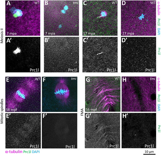
Fig. 7 Localization of Prc1l protein to microtubule structures involved in cytokinesis at the animal pole. (A-D′) Prc1l protein localizes to bundling midbody microtubules during meiosis II, (A,A′,C,C′) and becomes delocalized after polar body extrusion (D,D′). tmi mutants do not show antibody labeling (B,B′), as expected from antibody specificity (see also G,H and Fig. S9A). (E-H′) During embryonic mitotic divisions, Prc1l protein localizes to the microtubule apparatus (E,E′) and the FMA (G,G′), with the expected absence of label in tmi mutant embryos (F,F′,H,H′).
Acknowledgments
This image is the copyrighted work of the attributed author or publisher, and
ZFIN has permission only to display this image to its users.
Additional permissions should be obtained from the applicable author or publisher of the image.
Full text @ Development