
|
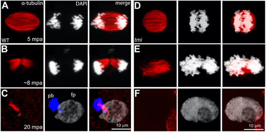
Fig. 5 Meiotic spindle defects in tmi mutants. (A-C) In wild-type eggs, spindles for the second meiotic division exhibit an elongated shape (A) that resolves into midbody-like structures (B) and becomes highly bundled during abscission (C). During the last stages of cytokinesis, the midbody becomes asymmetric with a longer region adjacent to the polar body (pb; left DNA mass in C, which becomes condensed), and a shorter region adjacent to the female pronucleus (fp; right DNA mass in C, which undergoes decondensation). (D-F) In eggs from tmi mutant females, spindles appear shorter with less-defined poles (D) and fail to form a well-defined midbody (E; see also Fig. 7B). Subsequently, a single DNA mass is observed (F), likely after failure of cytokinesis for meiosis II followed by fusion. In all panels, DNA is represented in white, except the polar body in C, which has been color-coded in blue in the appropriate z-stack sections to better highlight this structure.