
|
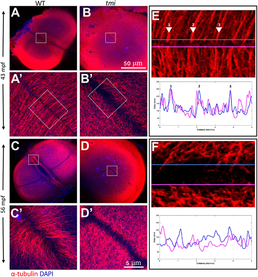
Fig. 4 Aberrant reorganization and clustering in the furrow microtubule array. Immunolabeling of fixed embryos to detect microtubules. (A,B) During early stages of furrow formation, the FMA forms along the length of the furrow, consisting of tubule bundles parallel to each other and perpendicular to the furrow (A,A′). tmi mutants fail to show a clearly organized FMA and exhibit a reduction in bundling (B,B′; see also Fig. S5). (C-D′) Upon furrow maturation, tubules of the FMA reorganize, becoming enriched to the distal ends of the furrow and acquiring a tilted angle pointing distally (C,C′), whereas in tmi mutants microtubules fail to reorganize in this manner (D,D′). (E,F) Line scans of FMA bundles along both sides of the furrow show a strong spatial concordance of microtubule bundles in wild type (E, representing the boxed area in A′; numbers correspond to major sites of bundling), consistent with interconnections between bundles across the furrow. Such spatial concordance is absent in tmi mutants (F, corresponding to the boxed area in B′). Images are representative of nine ROIs, each spanning the length of the furrow for each condition (see Fig. S5 and Materials and Methods). Boxed areas in A-D are shown at higher magnification in A′-D′, respectively.