
|
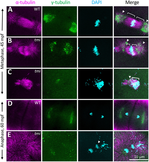
Fig. 3 Spindle formation defects in the early mitotic divisions in tmi mutants. (A-C) At metaphase, wild-type asters are highly symmetric (A). In tmi mutants, spindles are often asymmetric (B) or unipolar with chromosomes clustering at one end of the structure (C). Brackets and arrowheads highlight kinetochore microtubule and centrosomes, respectively. (D,E) During anaphase, tmi mutants exhibit chromosomes that fail to segregate (E, arrowhead), which was not observed in wild type (D). Analysis is from embryos at the two- to eight-cell stages (0.751-0.25 hpf), with the specific cell cycle stage assigned by chromosome and spindle morphology.