
|
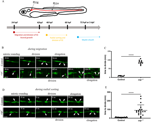
Fig. 1 SCs show a regular pattern of division during migration and radial sorting. (A) Timeline of SC behavior and myelination in zebrafish. SCs migrate and divide along growing axons between 24 hpf and 48 hpf. They start to radially sort axons of the PLLn at around 48 hpf and divide intensively. The myelin sheath was analyzed starting from 3 dpf (or 72 hpf). (B) Still images of time-lapse imaging in Tg(foxd3:gfp) control and Tg(foxd3:gfp)/csp−/− embryos at around 30 hpf. Arrows indicate SCs along the PLLn at different time points prior to and after division. Scale bar: 20 µm. (C) Quantification of the time required for control (11 cells from three different embryos) and csp−/− (ten cells from three different embryos) SCs to successfully complete cytokinesis during migration (****P≤0.0001). (D) Still images of time-lapse imaging in Tg(foxd3:gfp) control and Tg(foxd3:gfp)/csp−/− embryos at around 52 hpf. Arrows indicate SCs along the PLLn at different time points prior to and after division. Scale bar: 20 µm. (E) Quantification of the time required for control (20 cells from six different embryos) and csp−/− (20 cells from five different embryos) SCs to successfully complete cytokinesis (****P≤0.0001). m, minutes.