
|
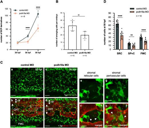
Fig. 4 Pcdh18a C-terminal truncation reduces the number of stromal cells. (A) Counting of ET37:GFP+ SCP derivatives in the developing ventral tail over a five-somite width at 26, 36 and 50 hpf. Plots represent biological replicates from a single experiment; n=6 for control and pcdh18a-ΔCP106 morphants (mean±s.d.; ****P<0.0001; unpaired, two-tailed Student's t-test). (B) Counts of the number of emerging SCPs within a five-somite width, from 24 to 40 hpf (from the same time-lapse confocal sequences as in Fig. 2C), n=6 embryos per condition (mean±s.d.; **P=0.0037; unpaired, two-tailed Student's t-test). (Ca-Cd) Confocal spinning-disk projections of live Tg(ET37:GFP; kdrl:ras-mCherry) control or pcdh18a-ΔCP106 embryos at 50 hpf. ca, caudal artery. Scale bars: 30 µm. (Ce-Ch) Dashed boxes in Cb indicate the regions enlarged in Ce-Ch, which show the two stromal cell types found in the CHT at 50 hpf. SRCs interconnect the vascular segments of the plexus and seem to maintain its architecture (Ce, and arrows in Cf), whereas SPvCs are spread along and around the vessels (Cg, and arrows in Ch). Scale bars: 10 µm. (D) Numbers of the three types of SCP derivatives – SRCs and SPvCs in the CHT, and FMCs in the caudal fin – in 14 embryos per condition, counted over a five-somite width (mean±s.d.; **P=0.0011; ****P<0.0001; unpaired, two-tailed Student's t-test).