
|
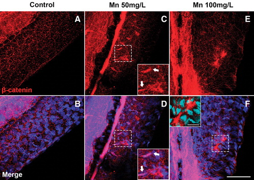
Fig. 5 Mn treatment disorganizes the epithelium and induces the expression of β-Catenin in the caudal fin. Confocal analysis of zebrafish larvae at 96 h postfertilization stained with β-Catenin (red) and DAPI (blue). In the control embryo, β-Catenin is diffused at typical intercellular adherens junctions of epithelial cells (A, B). In the 50 mg/L Mn-treated embryo (C, D), β-Catenin-positive intercellular junctions appear disorganized and into the nuclei of some mesenchymal cells. In embryos treated with 100 mg/L Mn (E, F), β-Catenin is concentrated in some areas of the epithelial tissue. Scale bar in F = 50 μm and in the inset = 25 μm. The groups were performed with n = 5. DAPI, 4,6-diamidino-2-phenylindole dihydrochloride. Color images are available online.