IMAGE

Figure 6

ID
ZDB-IMAGE-231211-22
Source
Figures for Ricci et al., 2023
Image
Figure Caption

Figure 6

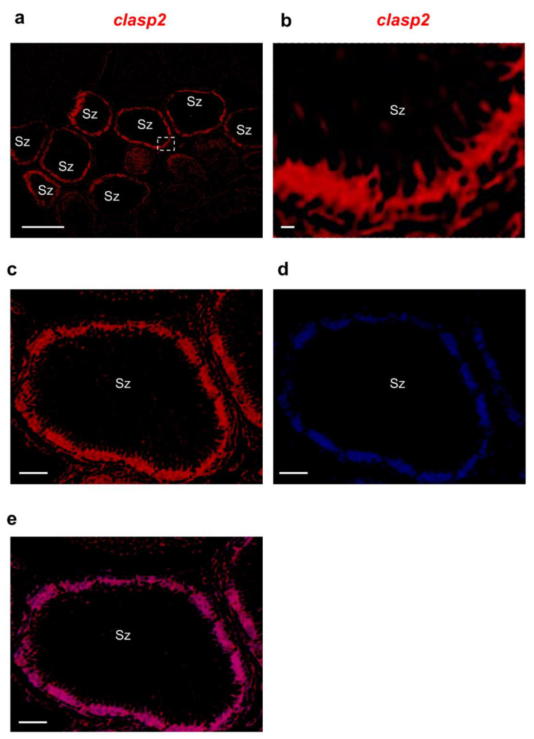
Clasp2 mRNA distribution in adult guppy testis, confocal microscopy. (a) Fluorescence in situ hybridization of clasp2 shows the labeling concentrated at the periphery of numerous spermatozeugmata. (b) High magnification of a portion of spermatozeugmata in a white rectangle is shown in panel a. (c) Fluorescence in situ hybridization of clasp2 is seen at the periphery of spermatozeugmata cysts. (d) DAPI staining marks all the cell nuclei of the testis. (e) The merge of fluorescence in situ hybridization of clasp2 and DAPI in adult guppy testis. Abbreviation: Sz; spermatozeugmata. Scale bars are 50 µ (a); 10 µ (b); and 30 µ (ce).

Acknowledgments
This image is the copyrighted work of the attributed author or publisher, and ZFIN has permission only to display this image to its users. Additional permissions should be obtained from the applicable author or publisher of the image. Full text @ Animals (Basel)