IMAGE

Figure 1

ID
ZDB-IMAGE-231205-1
Source
Figures for Wilcockson et al., 2023
Image
Figure Caption

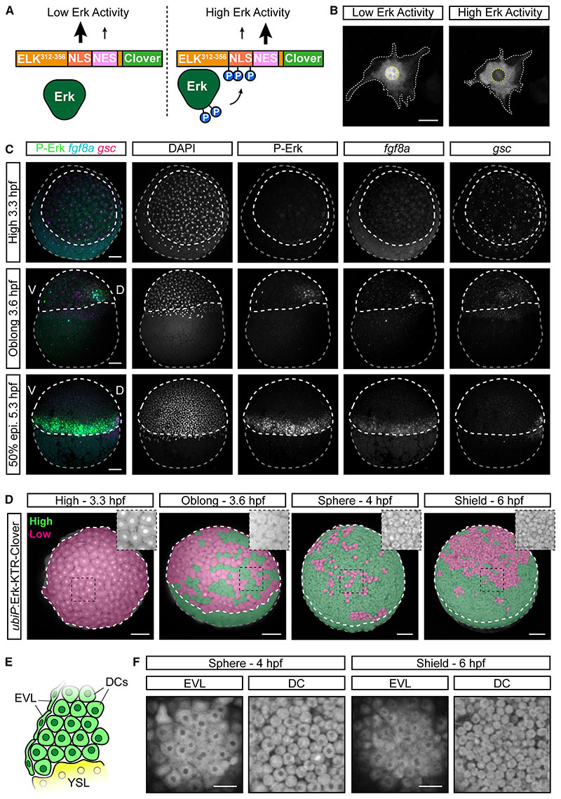
Figure 1 Off-target Erk-KTR activity in the early zebrafish embryo

(A) Schematic of the Erk-KTR construct showing the N-terminal Erk-docking domain derived from ELK1, a nuclear localization sequence (NLS) containing Erk-consensus phosphorylation sites (P), a nuclear export sequence (NES) and a C-terminal fluorescent protein, Clover.

(B) Live images of an NIH-3T3 cell transfected with ubiP:Erk-KTR-Clover construct. The cytoplasmic-to-nuclear ratio of the Erk-KTR fluorescence provides a live readout of relative Erk activity levels. White-dashed line, cytoplasm; yellow-dashed line, nucleus.

(C) Combined immunofluorescence and RNAscope showing diphosphorylated Erk (P-Erk) and fgf8a expression relative to the dorsal organizer marked by goosecoid (gsc) expression. Embryos are oriented in an animal view (3.3 hpf) or lateral view (3.6-5.3 hpf). White-dashed line, embryo proper; gray-dashed line, yolk; 50% epi, 50% epiboly.

(D) Stills of live ubiP:Erk-KTR-Clover embryos. Embryos are false-colored to indicate Erk-KTR activity as readout by the KTR reporter in a binary manner (green, high activity; magenta, low activity). Embryos are shown from an animal-lateral view. Insets show a magnified view of the boxed region without false coloring.

(E) Schematic cross-section of the embryonic margin showing the relative position of the deep cells (DCs), the enveloping layer (EVL) and the yolk syncytial layer (YSL).

(F) Single z-slices showing Erk-KTR activity in the EVL and DCs from the indicated embryos in (D).

Scale bars, 25 μm (B), 50 μm (F), or 100 μm (C and D).

Acknowledgments
This image is the copyrighted work of the attributed author or publisher, and ZFIN has permission only to display this image to its users. Additional permissions should be obtained from the applicable author or publisher of the image.

Reprinted from Developmental Cell, 58(23), Wilcockson, S.G., Guglielmi, L., Araguas Rodriguez, P., Amoyel, M., Hill, C.S., An improved Erk biosensor detects oscillatory Erk dynamics driven by mitotic erasure during early development, 2802-2818.e5, Copyright (2023) with permission from Elsevier. Full text @ Dev. Cell