IMAGE

Figure 1

ID
ZDB-IMAGE-231127-26
Source
Figures for Marines et al., 2023
Image
Figure Caption

Figure 1

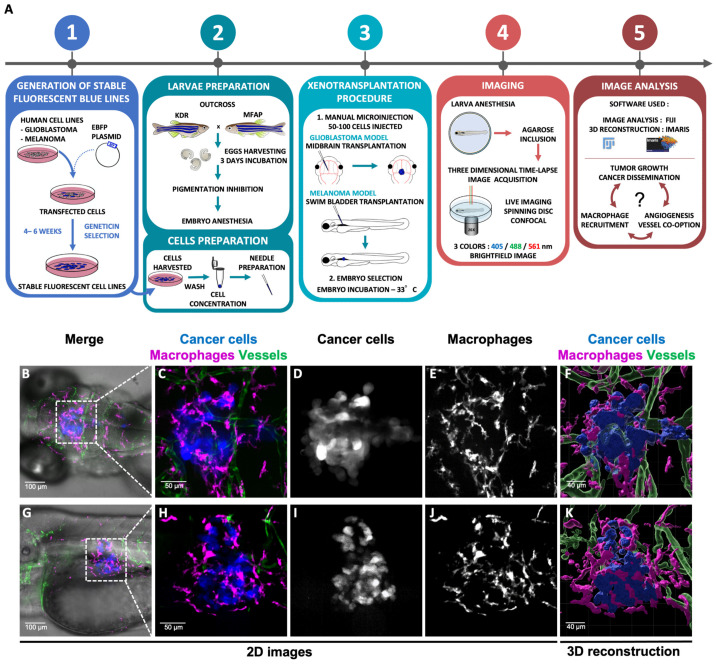
Experimental workflow and 3D glioblastoma and melanoma TME reconstruction. (A). The method is composed of five successive steps. Blue glioblastoma (GBM) and melanoma fluorescent human cancer cell lines (U87 and A375, respectively) were firstly generated (1). For this purpose, GBM cells and melanoma cells were transfected with a EBFP2 plasmid and selected over several weeks using Geneticin (1). Then, cells and embryos were prepared prior to transplantation (2). GBM cells were transplanted in the midbrain and melanoma cells in the swim-bladder of 3 dpf embryos (3). Imaging and time lapse were performed at 24 h or 48 h post transplantation (4). Image analysis was achieved using Fiji and Imaris software (5). (BK) Each cell population is highlighted with a coded colour; cancer cells in blue or white, macrophages in magenta or white and vessels in green. (BE) Visualization of GBM TME in the midbrain of zebrafish embryo at 4 dpf and 24 h post transplantation. Images represent a Z projection of 67 slices. (GJ) Visualization of Melanoma TME in the swim bladder of zebrafish embryo at 4 dpf and 24 h post transplantation. Images represent a Z projection of 74 slices. (F,K) A 3D reconstruction of tumours infiltrated by macrophages and vessels. (E,J) A high number of macrophages are recruited all around the tumour sites.

Acknowledgments
This image is the copyrighted work of the attributed author or publisher, and ZFIN has permission only to display this image to its users. Additional permissions should be obtained from the applicable author or publisher of the image. Full text @ Curr. Iss. Mol. Biol.