IMAGE

Fig. 5

ID
ZDB-IMAGE-231124-33
Source
Figures for Saunders et al., 2023
Image
Figure Caption

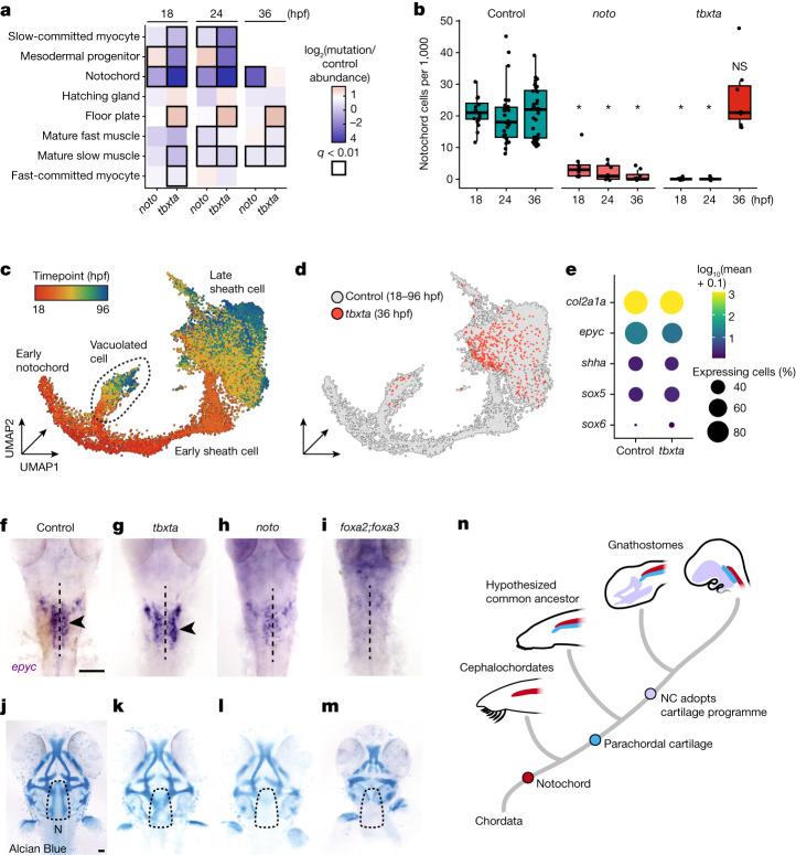
Fig. 5 Tbxta and Noto perturbations uncover the genetic requirements of cranial cartilage development.

a, Axial and paraxial mesodermal derivatives and their cell abundances relative to control embryos at three timepoints for tbxta and noto crispants. Black squares indicate significance (q < 0.01, beta-binomial regression). b, Box plots of notochord cell counts from individual embryos for controls, noto and tbxta crispants. Significance (*q < 1 × 10−5) is relative to wild-type control-injected embryos. Thick horizontal lines represent medians, box edges delineate first and third quartiles, respectively and whiskers extend to ±1.5× interquartile range. c, UMAP embedding of the notochord trajectory constructed with reference cells and tbxta cells. Cells are coloured by timepoint and are labelled by subtype annotation. d, UMAP embedding of notochord cells, coloured by genotype. e, A dotplot for a subset of genes that are expressed in notochord sheath, and in tbxta-independent cells, which are referred to as NLCs in the text. Colour represents mean normalized gene expression, and circle size indicates the percentage of notochord cells expressing the gene at 36 hpf. fi, epyc ISH (36 hpf; dorsal, anterior view) in control (f), tbxta (g), noto (h) and foxa2;foxa3 (i) crispants. The dashed line indicates the notochord, and parachordal cartilage cells in control and tbxta crispants are marked by black arrowheads. Scale bar, 100 µm. jm, Alcian Blue staining of 72 hpf control (j), tbxta (k), noto (l) and foxa2;foxa3 (m) crispants. Dashed outline surrounds the parachordal cartilage region. All tbxta, noto and foxa2/foxa3 crispants lack a notochord. (N, notochord; dotted line surrounds the parachordal cartilage). Scale bar, 100 µm. n, A model depicting the hypothesized relationship between the notochord (NC) and cranial cartilage and bone elements over chordate evolution.

Source Data

Acknowledgments
This image is the copyrighted work of the attributed author or publisher, and ZFIN has permission only to display this image to its users. Additional permissions should be obtained from the applicable author or publisher of the image. Full text @ Nature