IMAGE

Fig 6

ID
ZDB-IMAGE-231118-7
Source
Figures for Veen et al., 2023
Image
Figure Caption

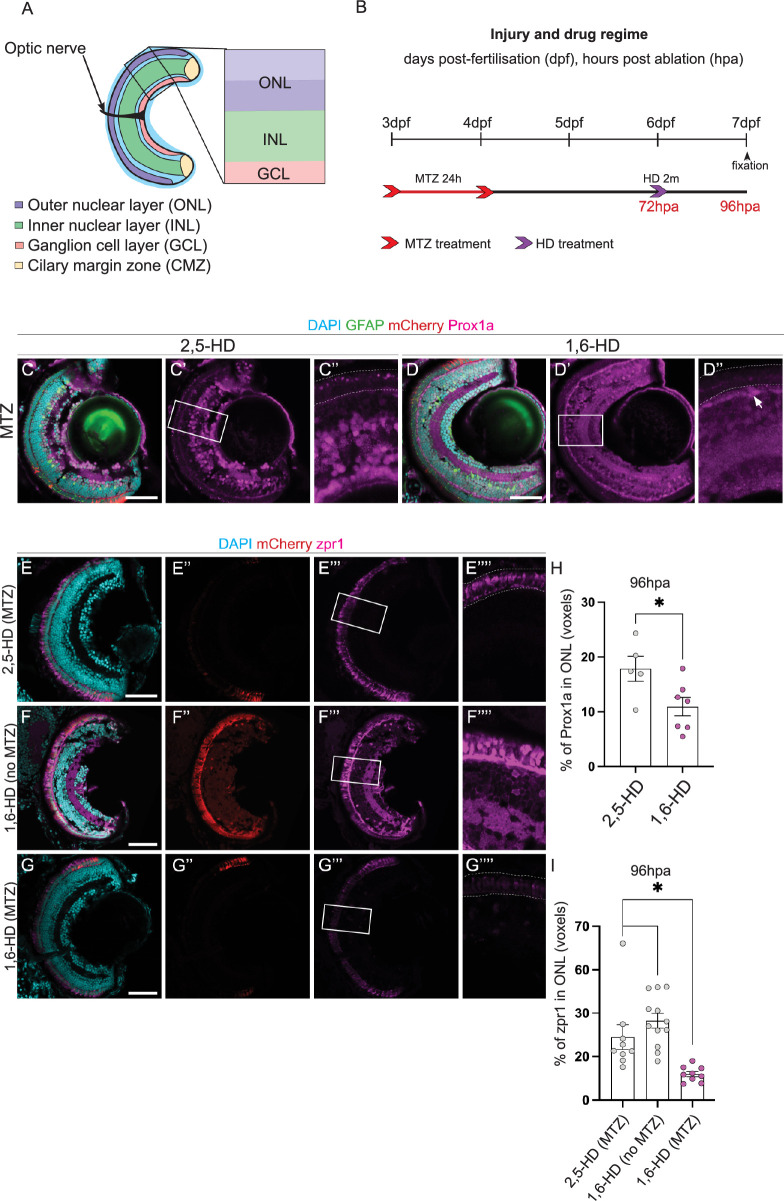
Fig 6 Prox1a puncta in liquid-liquid phase separates regulate PR differentiation.

(A) Schematic highlighting the retinal layers analysed in the zebrafish larvae. (B) Experimental timeline. Larvae were swum in metronidazole (MTZ) for 24 hours at 3dpf (days post-fertilisation), followed by a 2 min treatment with 1,6-Hexanediol (1,6-HD) or 2,5-HD at 6dpf, before processing at 7dpf. (C-D”) Following photoreceptor (PR) ablation, Tg(lws2:nfsb-mCherry, gfap:eGFP) retinas show Prospero homeobox1a (Prox1a, pink) puncta only in the ONL of 2,5-HD control treated retinas (n = 5, 17.86 ±2.3), but not when liquid-liquid phase separates have been disassembled in the 1,6-HD treated samples (n = 7, 10.94 ±1.681, quantified in H. Arrow indicates nuclear Prox1a staining outside of the photoreceptor layer. (E-G”’) zpr1 cone photoreceptors are observed in both control samples (2,5-HD treated ablated retinas and 1,6-HD treated, non-ablated retinas), but much reduced in the ONL of 1,6-HD treated ablated retinas, quantified in I. DAPI labels cell nuclei (blue). Scale bars: 50 μm.

Acknowledgments
This image is the copyrighted work of the attributed author or publisher, and ZFIN has permission only to display this image to its users. Additional permissions should be obtained from the applicable author or publisher of the image. Full text @ PLoS Genet.