
|
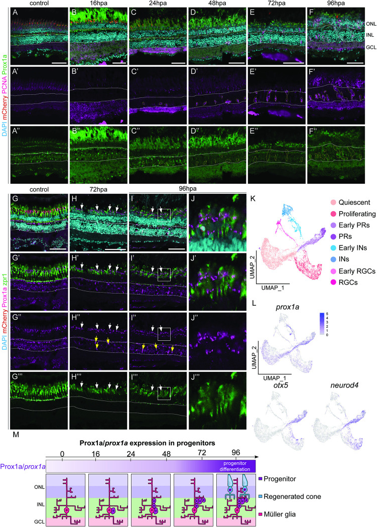
Fig 4 Prox1a is expressed in the photoreceptors within the outer nuclear layer.
(A-F”) Within adult retinas in uninjured control, post ablation (hpa), Prospero homeobox1a (Prox1a, green) is expressed in the inner nuclear layer (solid white outline), but never in proliferating (proliferative cell nuclear antigen positive, PCNA, pink) cells (dotted outlines). At 72hpa and 96hpa Prox1a in addition expressed in the ONL. (G-I”) cone photoreceptor marker, zpr1 (green) is expressed in the ONL in the control and at all the time points post ablation. (J-J”’) is the magnified images of (I-I”’). At 72hpa and 96hpa, Prox1a is expressed as distinct puncta within zpr1 expressing photoreceptors (arrows and high-power inset). Scale bars: 50 μm. (K, L) UMAP plot of FACS MG 3 days after injury can be subdivided into distinct MG derived cell population based on stereotypical markers. The feature plots show that