IMAGE

Fig 1

ID
ZDB-IMAGE-231118-1
Source
Figures for Veen et al., 2023
Image
Figure Caption

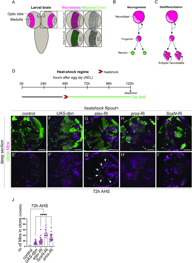
Fig 1 Knockdown of Dpn, Elav, Pros, and SoxN result in dedifferentiation of medulla neurons.

(A) Schematic representation of Drosophila larval central nervous system (CNS). The medulla (pink) is located with the Optic Lobes (OLs) of the CNS. The superficial surface of the medulla (0μm) contains neuroblasts (NBs, pink), whereas the deep layers (8–12μm) contain mature neurons (green). A single NB in the superficial layer and its neuronal progeny in the deep layers are labelled in light green. (B) During neurogenesis NBs undergo asymmetric divisions to generate a NB and an intermediate progenitor (ganglion mother cell, GMC). The GMC produces 2 neurons. (C) During dedifferentiation, mature neurons dedifferentiate to give rise to ectopic NBs, which continue to divide and create tumours. (D) Experimental design to induce clones with manipulate gene expression. Clones induced via heat shock (red arrows) at 48 hours after egg lay (AEL) are dissected 72 hours after heat shock (AHS). (E-I’) In the deep layers of the medulla, control clones contained no stem cells (E, E’), clonal overexpression of UAS-dpn (deadpan), elav-Ri (embryonic lethal), pros-Ri (prospero), SoxN-Ri (SoxNeuro) resulted in stem cell induction (Miranda+, magenta) in the deeper mature neuron layers, indicative of dedifferentiation (J). (J) Quantification of % Mira+ cell volume vs. clone volume: Control (n = 9, 1.874 ±0.4897), UAS-dpn (n = 17, 33.62 ±3.243), elav-Ri (n = 22, 13.16 ±1.164), SoxN-Ri (n = 19, 21.74 ±2.926), pros-Ri (n = 11, 49.34 ±4.36). Data are represented as mean ± SEM. ****p < 0.0001. Scale bars: 50 μm. Deadpan data is adapted from [24].

Acknowledgments
This image is the copyrighted work of the attributed author or publisher, and ZFIN has permission only to display this image to its users. Additional permissions should be obtained from the applicable author or publisher of the image. Full text @ PLoS Genet.