Image
Figure Caption

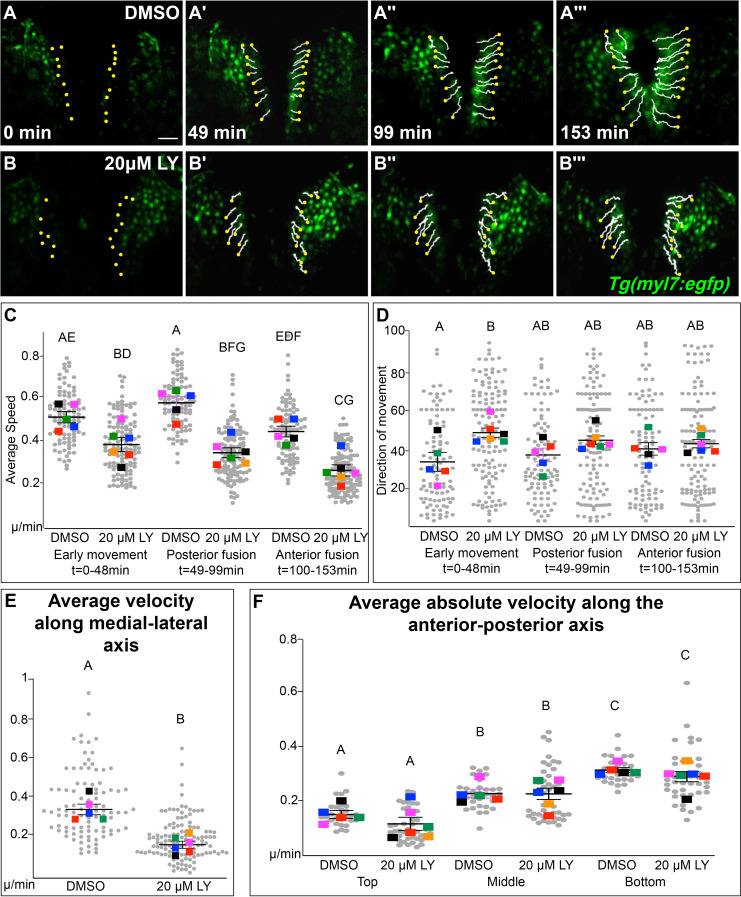
Figure 3—figure supplement 2. Phosphoinositide 3-kinase (PI3K) signaling directs myocardial movement during the early stages of cardiac fusion and regulates velocity along the medial-lateral axis.

(A, B) Time-lapse confocal reconstructions from Figure 3 overlaid with cell movement tracks, starting at t = 0 (yellow dots). Graphs of average speed (C) and direction of movement (D) subdivided into three distinct stages of cardiac fusion: early movement (0–48min), posterior fusion (49–99min), and anterior fusion (100–153min). Graphs of average absolute medial–lateral velocity (E) and average absolute angular velocity (F) sub-divided into cells at the top, middle, and bottom of the myocardial populations. The average speed of myocardial cells in LY-treated embryos is consistently slower than DMSO-treated embryos (C). This defect appears to derive mostly from differences in medial-lateral movement (E) rather than in angular movement (F). In LY-treated embryos, myocardial cells display a more angular average direction of movement compared to DMSO-treated embryos during the early stage of cardiac fusion (early movement), after which wild-type myocardial cell movement becomes more angular matching myocardial cell movements in LY-treated embryos (D). Gray dots – individual cells, color squares – average per embryo. Average and standard error based on embryos. Scale bar, 60μm. Letter change indicates p < 0.05, one-way analysis of variance (ANOVA) (C, D, F) or t-test (E). Raw data and full p-values included in the source file.

Acknowledgments
This image is the copyrighted work of the attributed author or publisher, and ZFIN has permission only to display this image to its users. Additional permissions should be obtained from the applicable author or publisher of the image. Full text @ Elife