
Image
|
Figure Caption
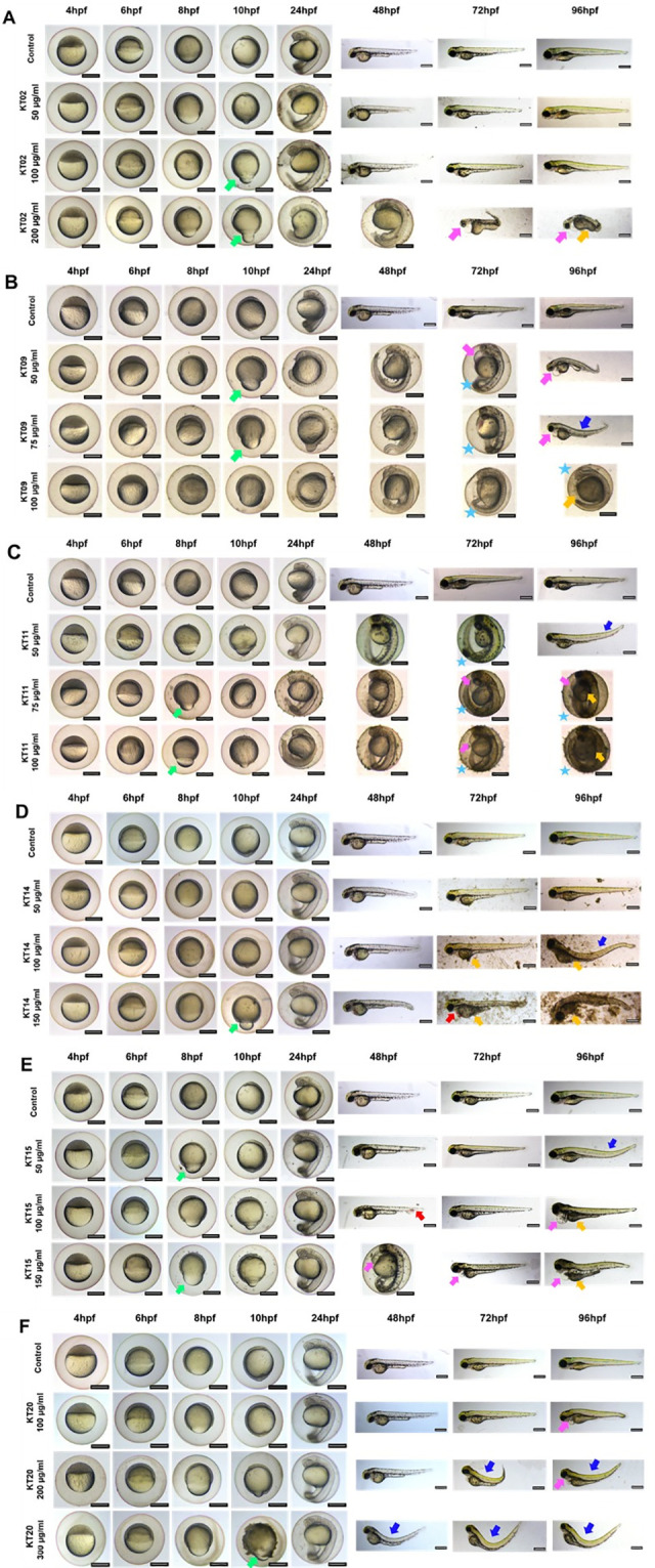
Fig 2 Morphological defects at different time points of exposure.
A-F: Representative images of 4–96 hpf larvae exposure to 6 plant extracts at the indicated concentrations. The morphology of embryos was observed and captured under a stereomicroscope. Scale bars = 400 μm. Green arrows indicate dumb-bell shape yolk, blue arrows indicate dorsal curvature, pink arrows indicate yolk sac edema, red arrows indicate hemorrhage orange arrows indicated necrosis, and stars indicate delayed hatching.
Acknowledgments
This image is the copyrighted work of the attributed author or publisher, and
ZFIN has permission only to display this image to its users.
Additional permissions should be obtained from the applicable author or publisher of the image.
Full text @ PLoS One