
|
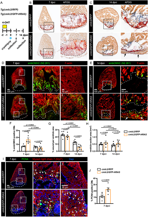
Fig. 7 Short-term expression of oncogenic HRAS accelerates heart regeneration. (A) Experimental design and transgenic fish lines. One pulse of 4-OHT was given 2 days before cryoinjury. Hearts were collected at 7 and 14 dpci. (B,C) AFOG staining to visualize myocardium in orange, fibrin in red and collagen in blue. The cryoinjured area is outlined with a dashed line. At 14 dpci, a new myocardial layer forms around the wound margin, as indicated by arrows. (D,E) Immunostaining for embCMHC (N2.261 antibody) to detect immature cardiomyocytes, co-stained with F-actin. At 14 dpci, a myocardial ‘bridge’ spanning the continuity of the ventricular wall is indicated by arrows (four hearts out of eight). (F-H) Quantification of the data representatively shown in D and E. cmlc2/RFP at 7 and 14 dpci, n=6; cmlc2/GFP-HRAS at 7 dpci, n=6, cmlc2/GFP-HRAS at 14 dpci, n=8; error bars indicate s.e.m.; two-way ANOVA with Šidák multiple comparisons test. (I) Immunostaining against PCNA to detect proliferating cardiomyocytes, labeled with Myl7. n=6. Some proliferating cells are indicated with arrowheads. (J) Quantification of data representatively shown in I. n=6. Data are mean±s.e.m.; unpaired two-tailed t-test.