IMAGE

Figure 2

ID
ZDB-IMAGE-231105-3
Source
Figures for Yilmaz et al., 2023
Image
Figure Caption

Figure 2

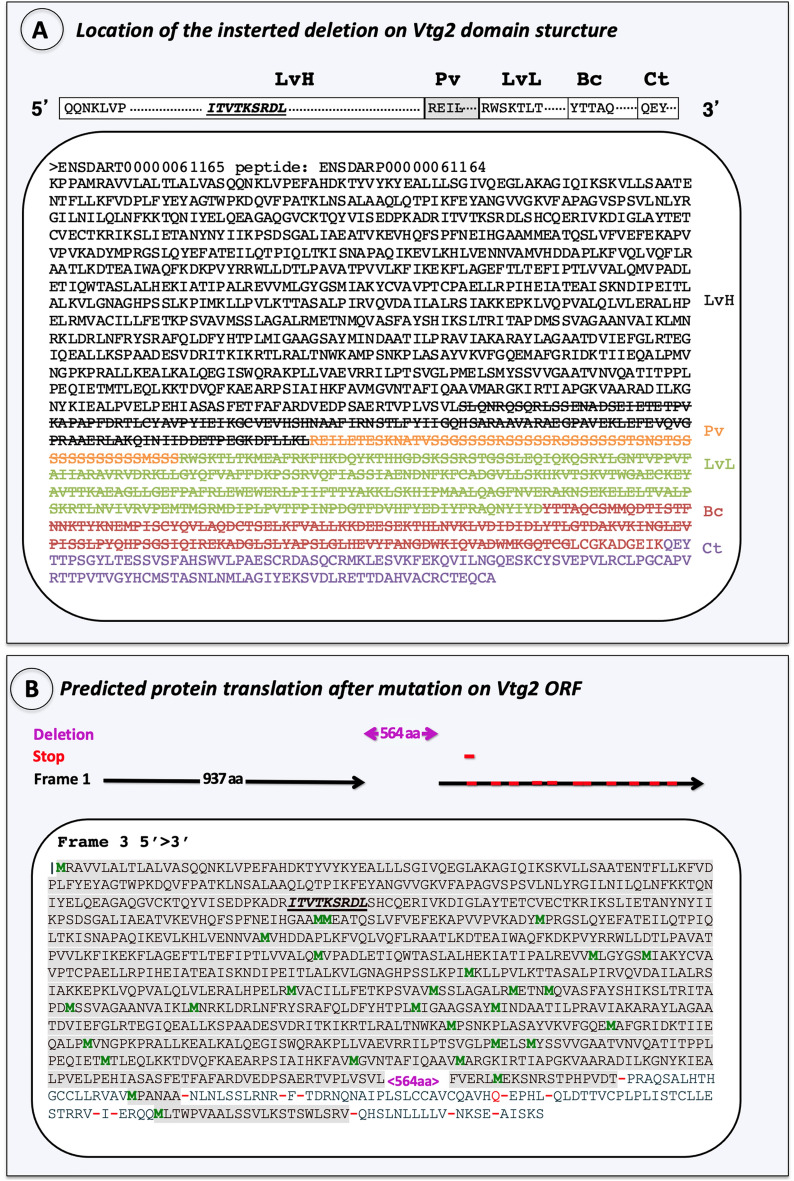
Characterization of the introduced mutation. (A) Location on Vtg2 polypeptide structure. The yolk protein domain structure of Vtg2 is pictured in 5′ > 3′ orientation above panel. Horizontal bars represent the heavy and light chain lipovitellins (LvH, LvL), phosvitin (Pv), beta component (Bc), and C-terminal (Ct) domains of the Vtg2 and are labeled above in large bold type. Sequences within these bars indicate the N-terminus of each yolk protein domain, the starting points of which are also indicated by shade color change to the polypeptide sequence shown below. Each domain is labeled to the right with the corresponding shade color. LvH: black, Pv: orange, LvL: light green, Bc: red, Ct: purple. Cas9 created mutations (large deletions) are indicated with stroked letters representing amino acid (aa) residues. (B) Frameshift caused by the introduced mutation on the Vtg2 open reading frame. A total of 973 aa which were not altered by the introduced mutation are indicated in grey shaded letters. The introduced mutation of 564 aa is indicated in magenta font set (<564 aa>). Methionine residues are shown in green font set and “stop codons” are indicated by red dashes. The receptor binding site is indicated by bold, italic and, underlined aa motif.

Acknowledgments
This image is the copyrighted work of the attributed author or publisher, and ZFIN has permission only to display this image to its users. Additional permissions should be obtained from the applicable author or publisher of the image. Full text @ Sci. Rep.