IMAGE

Fig. 6

ID
ZDB-IMAGE-231031-29
Source
Figures for Ka et al., 2023
Image
Figure Caption

Fig. 6

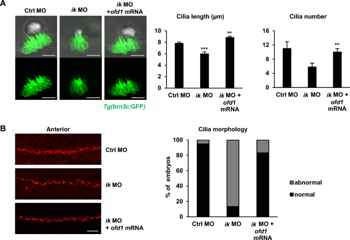
ofd1 co-injection can rescue ciliary dysmorphology of ik morphants (A) Confocal microscopic images of control MO, ik MO, and ik MO/ofd1 mRNA co-injected Tg(brn3c:GFP) embryos at 2 dpf. Scale bar, 20 µm. Bar graphs show quantification of ciliary length and number in the inner ears of control MO, ik MO, and ik MO/ofd1 mRNA co-injected embryos at 2 dpf. **p < 0.01, ***p < 0.001 (B) Whole-mount immunostaining of pronephric cilia using anti-acetylated-α-tubulin (red) at 2 dpf control MO, ik MO, and ik MO/ ofd1 mRNA co-injected embryos. Scale bar, 20 µm. Stacked bar graph displays the percentage of ciliary morphology of embryos in the anterior pronephric duct (percentage of abnormal ciliary morphology of embryos: control MO, 5.12% (37/39); ik MO, 86.6% (4/30); ik MO + ofd1 mRNA, 16.6% (15/18)

Acknowledgments
This image is the copyrighted work of the attributed author or publisher, and ZFIN has permission only to display this image to its users. Additional permissions should be obtained from the applicable author or publisher of the image. Full text @ Cell Biosci.