IMAGE

Figure 3

ID
ZDB-IMAGE-231030-120
Source
Figures for Huttner et al., 2023
Image
Figure Caption

Figure 3

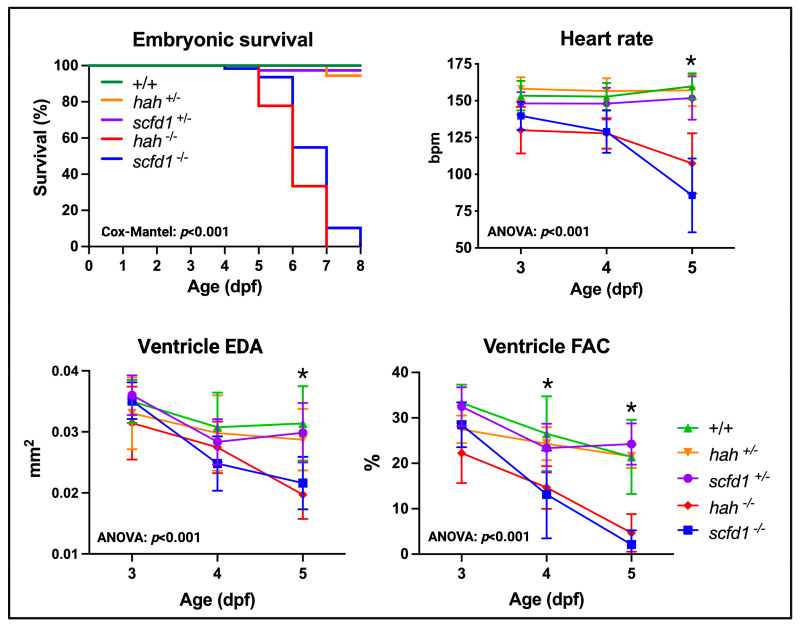
Scfd1 deficiency leads to cardiac dysfunction in zebrafish embryos. Survival, heart rate, ventricular end-diastolic area (EDA) and fractional area change (FAC) are reduced in embryonic hahvcc43−/− and scfd1vcc44−/− mutants, while heterozygous siblings of both lines (+/−) are indistinguishable from wildtype (WT; +/+). Non-parametric survival test (Cox-Mantel) and two-way ANOVA p-values stated. Animal numbers, mean values, and multiple comparison p-values for all variables, time points, and genotypes listed in Supplementary Table S2; asterisk indicates significant differences from WT in hahvcc4 −/− and scfd1vcc44−/− mutants. Significance if p < 0.05. Created with BioRender.com.

Acknowledgments
This image is the copyrighted work of the attributed author or publisher, and ZFIN has permission only to display this image to its users. Additional permissions should be obtained from the applicable author or publisher of the image. Full text @ J Cardiovasc Dev Dis