
Fig. 1
- ID
- ZDB-IMAGE-231020-1
- Publication
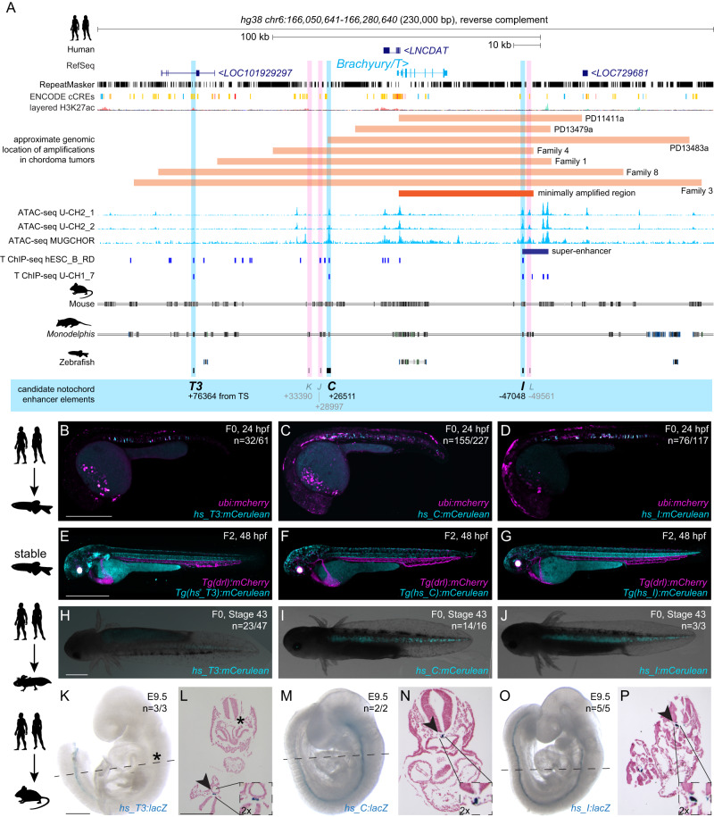
- Kemmler et al., 2023 - Conserved enhancers control notochord expression of vertebrate Brachyury
- All Figures
- Figures for Kemmler et al., 2023
|
Fig. 1
Human