IMAGE

Fig 3

ID
ZDB-IMAGE-231013-4
Source
Figures for Sakata et al., 2023
Image
Figure Caption

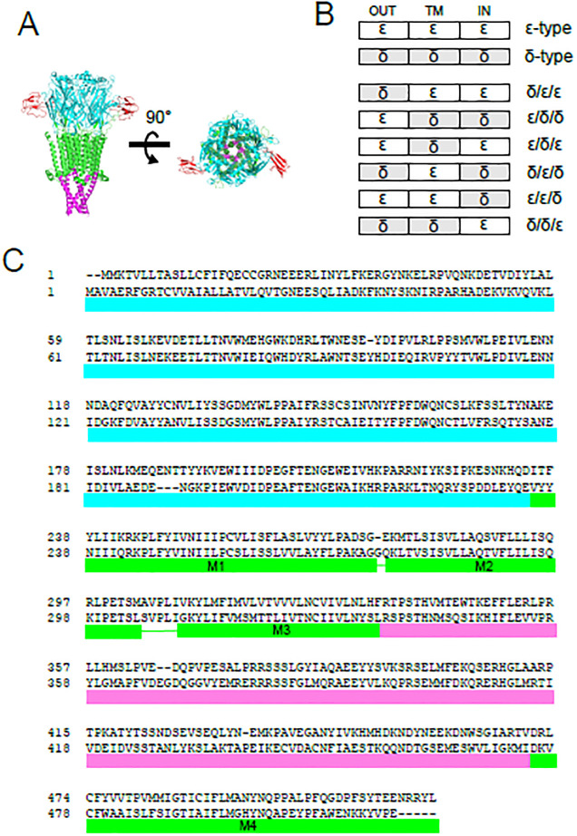
Fig 3 Chimeric subunits between the δ and the ε subunits.

(A), 3D structure of nAChR. The extracellular domain, the transmembrane domain and the intracellular domain are shown in cyan, green and magenta, respectively. Two bungarotoxin (BTX) molecules, shown in red, bind to the extracellular domain (PDB ID; 6uwz) [20]. (B), Schema of chimeras generated in this study. “OUT”, “TM”, “IN” represent the extracellular, transmembrane and intracellular domain, respectively. (C), Definition of the extracellular, transmembrane and intercellular domain in this study. Amino acid alignments of the upper lines and the lower lines are the δ and ε subunits, respectively. Cyan, green, magenta lines indicate the extracellular, the transmembrane and the intracellular domains, respectively.

Acknowledgments
This image is the copyrighted work of the attributed author or publisher, and ZFIN has permission only to display this image to its users. Additional permissions should be obtained from the applicable author or publisher of the image. Full text @ PLoS One