IMAGE

Fig. 4.

ID
ZDB-IMAGE-231007-4
Source
Figures for Bui et al., 2023
Image
Figure Caption

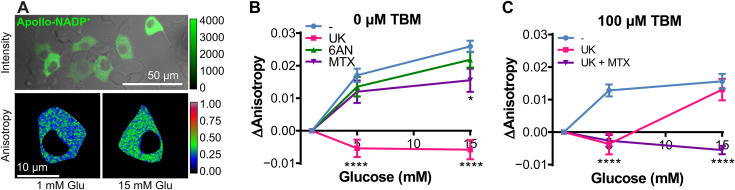
Fig. 4. Selective inhibition of NADP+ reduction pathways in unstressed and stressed INS1E pancreatic β cells.

(A) INS1E β cells transfected with Apollo-NADP+. Top: Fluorescence intensity image of Apollo-NADP+ expressed in INS1E β cells. Bottom: Fluorescence anisotropy images of Apollo-NADP+ expressing INS1E β cells stimulated with glucose. (B) Glucose-stimulated NADP+ reduction of INS1E β cells transfected with Apollo-NADP+. UK (50 μM) was used to inhibit pyruvate cycling, 6AN (5 μM) was used to inhibit the pentose phosphate pathway, and MTX (5 μM) was used to inhibit folate cycling. Chemical inhibitors were added 1 hour before imaging. (C) Glucose-stimulated NADP+ reduction of INS1E β cells transfected with Apollo-NADP+. Stress was induced by 8 hours of stress treatment with 100 μM TBM. n = 3 to 6 replicates. *P < 0.05 and ****P < 0.0001.

Acknowledgments
This image is the copyrighted work of the attributed author or publisher, and ZFIN has permission only to display this image to its users. Additional permissions should be obtained from the applicable author or publisher of the image. Full text @ Sci Adv