IMAGE

Fig 7

ID
ZDB-IMAGE-231005-10
Source
Figures for Unterweger et al., 2023
Image
Figure Caption

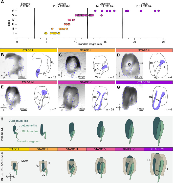
Fig 7 Ventral liver lobe formation during postembryonic growth.

(A) The 6 steps of ventral liver lobe formation correlate with fish standard length (SL). The numerical values that were used to generate the graph can be found in S1 Data. (B) Stage I: A small tissue extension at the tip of the left lobe is visible (n = 12 livers). (C) Stage II: a thin ventral lobe originates in the lower half of the left lobe (n = 6 livers). (D) Stage III: the thin ventral lobe shifts position towards the more anterior part of the left lobe (n = 4 livers). (E) Stage IV: the tip of the ventral lobe starts to expand (n = 7 livers). (F) Stage V: lateral-oriented expansion of the ventral lobe (n = 28 livers). (G) Stage VI: enlargement of all lobes in width (n = 6 livers). The blue areas in the schematics mark the region characteristic for the respective stage. (H) Schematic depicting the morphology of the liver in relation to the folding of the intestine in stages I-VI. A, anterior; P, posterior; R, right; L, left; RL, right lobe; LL, left lobe; VL, ventral lobe.

Acknowledgments
This image is the copyrighted work of the attributed author or publisher, and ZFIN has permission only to display this image to its users. Additional permissions should be obtained from the applicable author or publisher of the image. Full text @ PLoS Biol.