IMAGE

Fig. 4

ID
ZDB-IMAGE-231002-88
Source
Figures for Anderson et al., 2023
Image
Figure Caption

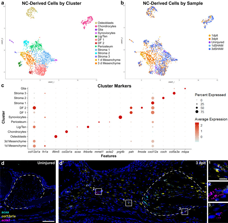
Fig. 4 Neural crest-lineage single-cell transcriptomics in early ligament regeneration.

a, b UMAP of single-cell RNA sequencing data of neural crest-derived cells from early ligament regeneration colored by (a) cluster or (b) sample. c DotPlot of marker genes used to identify each cluster. d RNAscope single-molecule fluorescent in situ hybridization (smFISH) for scxa (cyan), col12a1a (yellow), and acta2 (magenta) in an uninjured ligament. d’ smFISH for scxa, col12a1a, and acta2 expression at 3 dplt. Insets highlighting scxa+ col12a1a+ regenerative mesenchyme (inset 1, n = 5 per time point), acta2+ perivascular cells (inset 2), and acta2-low mesenchyme near the ligament stub (inset 3). Scale bars = 50 μm (d, d’), 10 μm (insets in d), ligament stub outlined in white dashed line.

Acknowledgments
This image is the copyrighted work of the attributed author or publisher, and ZFIN has permission only to display this image to its users. Additional permissions should be obtained from the applicable author or publisher of the image. Full text @ NPJ Regen Med