IMAGE

Fig. 3

ID
ZDB-IMAGE-231002-59
Source
Figures for Anderson et al., 2023
Image
Figure Caption

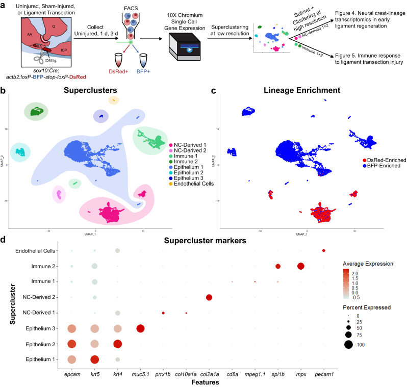
Fig. 3 Single-cell transcriptomics of joint tissue through early ligament regeneration.

a Schematic detailing the generation and analysis of single-cell RNA sequencing data. Jaw joints from uninjured, 1 day post-SHAM (nicking of the epithelium above the ligament) surgery, 3 days post-SHAM surgery, 1 day post-ligament transection, and 3 days post-ligament transection were dissociated and sorted for DsRed (neural crest-derived) or BFP (non-neural crest-derived). b UMAP of low-resolution superclusters for merged samples. Superclusters include 2 neural crest-derived clusters (NC-Derived), 2 immune clusters, 3 epithelial clusters, and 1 endothelial cluster. c UMAP colored for neural crest-lineage enrichment. Cells from FACS-sorted DsRed+ neural crest-derived cells colored in red, cells from sorted BFP+ non-neural crest-derived cells colored in blue. d DotPlot detailing expression of marker genes for each supercluster.

Acknowledgments
This image is the copyrighted work of the attributed author or publisher, and ZFIN has permission only to display this image to its users. Additional permissions should be obtained from the applicable author or publisher of the image. Full text @ NPJ Regen Med