
|
Figure 4.
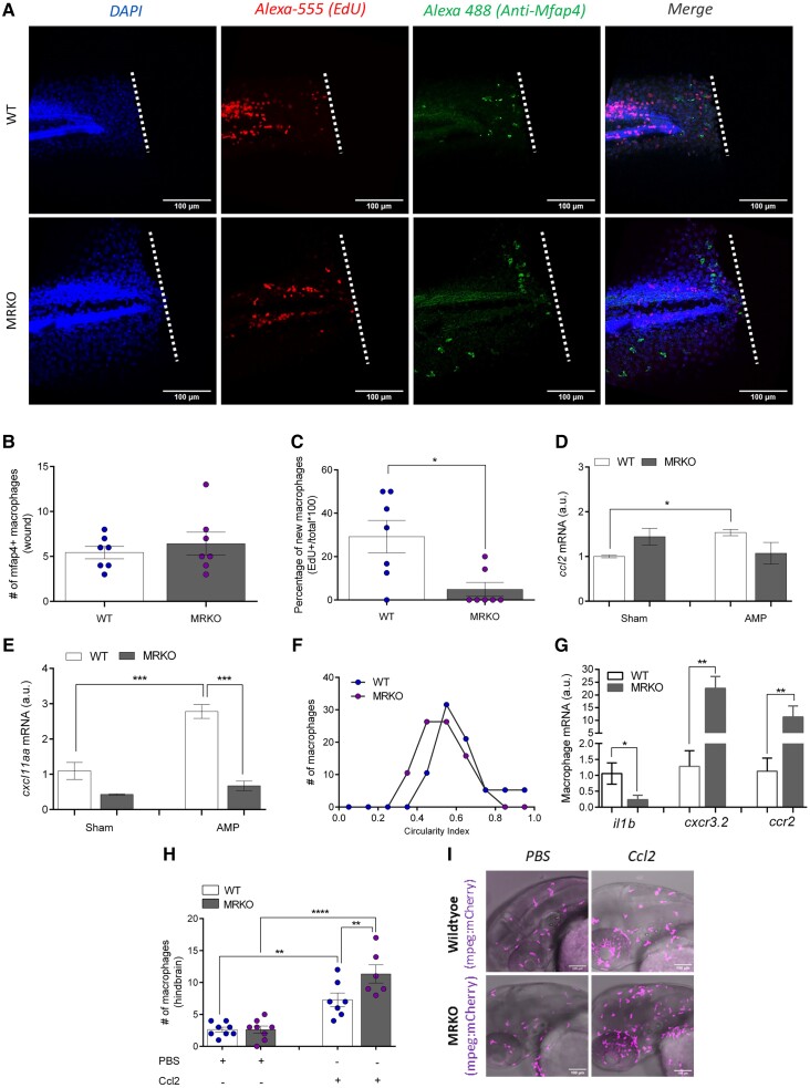
The generation of new macrophages is reduced in MRKO larvae, and macrophages migrating toward the wound exhibit a less inflammatory phenotype. (A) Representative images of macrophage proliferation at 48 hours postfertilization (hpf), 4 hours postamputation (hpa), where the nuclei are stained with DAPI (blue), new cells have incorporated the thymidine nucleoside analogue EdU (red), and macrophages are visualized by immunohistochemistry using a Mfap4 antibody (green). White dotted lines denote the location of amputation. (B) Number of Mfap4+ macrophages localized to the wound site at 4 hpa (n = 7). (C) The percentage of new (EdU+) macrophages localized to the wound site at 4 hpa (n = 7). (D and E) Transcript abundance of macrophage-specific chemokines