
Image
|
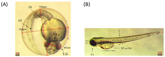
Figure Caption
Fig. 1 Inserts show a representative 48 hpf embryo (A) and 72 hpf larva (B), displaying the analyzed parameters. D1: Eye Diameter 1; D2: Eye Diameter 2; D.B.E.: Distance between eyes; E.D.: Eye diameter; E.L.: Eye length; Y.P.: Yolk perimeters; Y.A.: Yolk area; L: body length. Scale bar 100 µm.
Acknowledgments
This image is the copyrighted work of the attributed author or publisher, and
ZFIN has permission only to display this image to its users.
Additional permissions should be obtained from the applicable author or publisher of the image.
Full text @ Int. J. Mol. Sci.