
|
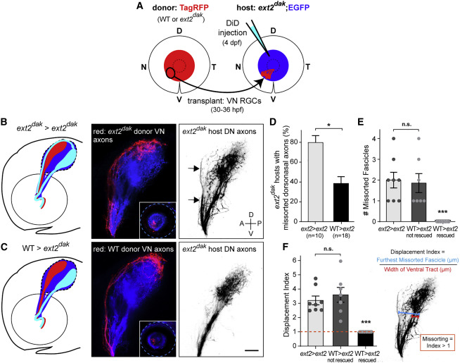
Fig. 1 HS-mediated trans-axonal signaling corrects pre-target topographic sorting errors
(A) Ventronasal (VN) RGCs of an isl2b:TagRFP donor embryo (WT or ext2dak mutant lacking HS) were topographically transplanted between 30 and 36 hpf into the VN retina of an isl2b:EGFP ext2dak mutant host. Pre-target sorting of host dorsonasal (DN) axons along the optic tract was assessed by injecting DiD into the host DN retina at 4 dpf.
(B and C) Diagrams and lateral views of donor VN axons (red) and host DN axons (inverted images) along the optic tract of ext2dak hosts at 4 dpf.
(B) Ext2dak DN axons missort along the dorsal branch of the optic tract in ext2dak hosts transplanted with ext2dak VN RGCs (arrows).
(C) Missorting of ext2dak DN axons is corrected after transplanting WT VN RGCs. Scale bar: 50 μm.
(D) Quantifications of the percentage of transplanted ext2dak hosts with missorted DN axons at 4 dpf. One-sided chi-squared test with Yates’s correction, X2 (1, N = 28) = 2.872, p = 0.0451.
(E) Number of missorted DN axon fascicles in ext2dak hosts with sorting defects. One-way ANOVA with Tukey’s post-hoc test, p ˂ 0.0005.
(F) Quantification of the displacement index in ext2dak hosts with sorting defects. One-way ANOVA with Tukey’s post-hoc test, p ˂ 0.0001. Error bars: standard errors.
See also Figure S1.